7th Meeting
Leiden, The Netherlands, April 21-23, 2008


Unexpected role of glycosphingo-lipids in cell physiology.
L1
G van Meer, J Wolthoorn, S Groux-Degroote, D van den Heuvel1, H Gerritsen1, D Halter, S Neumann, S van der Poel, H Sprong
Bijvoet Center and Institute of Biomembranes, 1Molecular Biophysics, Utrecht University, The Netherlands
Until a decade ago, the biosynthesis of the sphingolipids seemed simple: sphingosine, newly synthesized or recycled from the lysosomes, is converted to ceramide in the ER, where after vesicular transport it receives a phosphocholine or glucose headgroup in the cis-Golgi lumen. Now we know that there are at least 6 ceramide synthases, that sphingomyelin is synthesized in thetrans-Golgi, that the ceramide is supplied via the ceramide transfer protein CERT, and that CERT is regulated via PI4P on the Golgi and phosphorylation. Unexpectedly, glucosylceramide is synthesized on the cytosolic surface of the Golgi, and must cross the membrane to reach the site of complex glycolipid synthesis in the Golgi lumen.
We found that natural glucosylceramide does not cross the Golgi membrane and is not a substrate for the multidrug transporters ABCB1 and C1. Instead, glucosylceramide was transported back to the ER by a mechanism involving the glucosylceramide binding protein FAPP2 bound to the trans- Golgi via PI4P. It then crosses towards the ER lumen and reaches the Golgi lumen by vesicular transport, possibly generating ER sphingolipid domains along the way. Mean- while, glucosylceramide is required for protein sorting in the secretory pathway by affecting conditions in the lumen. As a possible explanation, we found that glucosylceramide affects the lumenal pH.
The (patho)physiological role of sphingolipids in vascular biology.
L2
SLM Peters, LJA Spijkers, RFP van den Akker, PB van Loenen, N Hajji and AE Alewijnse
Dept. Pharmacology & Pharmacotherapy, Meibergdreef 15, 1105 AZ Amsterdam, The Netherlands
Besides their well-documented effects on cellular growth, sphingolipids have vasoactive properties. Although sphingolipids are present in blood in high concentrations, both endothelial as well a vascular smooth muscle cells can synthesize the different sphingomyelin metabolites. Since these celle also express the different target molecules, sphingomyelin metabolites can be regarded as auto- or paracrine factors in the vascular system. Indeed, several vasoactive factors exert their vasoactive effects partly by modulation of sphingolipid metabolism in the vessel wall. For instance, both angiotensin II as wel as muscarinic receptor agonist can activate endothelial NO synthase by activation of sphingosine kinase. Interestingly, the effect of locally formed S1P proved to be vessel type dependent. Whereas in larger conduit vessels endothelial S1P production leads to NO synthase activation, in resistance vessels locally formed S1P has an inhibitory effect on the release or action of endothelium- derived hyperpolarizing factor. Hypertension is associated with marked alterations in sphingolipid homeostasis; Inhibition of sphingosine kinase induces major constrictions in isolated vessels of hypertensive rats. Interestingly, these contractions are mediat- ed by an endothelium-derived cyclooxygenase 1 product. Endothelial sphingolipid metabolism plays an important role in controlling the release or action of dilatory or contracting factors.
P38-mediated endothelial cell death after ionizing radiation is under the control of ceramide generation and membrane remodeling.
L3
C Niaudet1, S Bonnaud1, S Gouard1, MH Gaugler1,2, I Corre1, F Paris1
1Centre de Recherche en Cancérologie Nantes-Angers, Inserm U892, Nantes, France; 2IRSN, Fontenay aux Roses, France
Despite the involvement of ceramide and acid sphingomyelinase (aSM) in irradiated endothelial cells (EC) apoptosis, molecular mechanisms involved are poorly understood. MAPK p38 has been considered as a crucial actor in EC radiosensitivity. If p38 activation has been shown to be dependent of DNA damage induction in tumor models, no relation has been described between ceramide and p38 under genotoxic stress. In the present study, we highlighted a direct link as p38 phosphostain- ing detected in intestine’s EC from 15 Gy-irradiated mouse was not observed in intestine’s EC from irradiated aSM KO mouse. In HMEC-1 EC, adding exogenous C16-ceramide or bacterial SM induced raft microdomain reorganisation from a discrete pattern in the cell surface to large and polarised areas, followed by p38 activation and cell death. Similar membrane remodeling following ceramide generation was also observed in 15 Gy- irradiated ECs and this membrane remodeling is a pre-requisite to p38 phosphorylation. The two concomitant phenomena, i.e. ceramide-induced raft coalescence and p38 death-pathway, have been validated by use of drugs, such as desipramine (aSM inhibitor), and nystatin (cholesterol scavenger, disorganizing lipid raft formation), which inactivated p38 and the subsequent EC death.
Ceramide: a novel modulator of toxoplasma gondii survival?
L4
S Sonda, I Bottova, AB Hehl
Institute of Parasitology, University of Zurich, Winterthurerstrasse 266a, CH-8057 Zurich, Switzerland.
Toxoplasma gondii, the most versatile and widespread member of Apicomplexan parasites, is responsible for a disease of major medical and veterinary importance worldwide. Despite the impact of toxoplasmosis, there are few currently available treatments, often resulting in general side-effects. Interest in invertebrates, that sustain the diffusion permeability barriers of epithelia and the glial blood-brain barrier. Trafficking of junction components to the correct membrane domain is interlocked to the process of cell polarization and relies on the correct compartmental- ization of proteins as well as lipids in membranes. Little is known about the contribution of lipids to cell junction formation. We use Drosophila to study the role of lipids (in particular sphingolipids, glycosphingo- lipids, and phospholipids) in these cell biological processes. As a handle to the genetic study of lipids in this context, we investigate the contribution of lipid metabolism genes to the process of SJ formation. We screened mutants in sphingolipid,glyco-sphingolipid, and phospholipid metabolism genes using an established dye penetration assay, which probes the ‘tightness’ of the epithelial permeability barrier. We now proceed to assess the molecular, cellular and ultrastructural defects in the mutants with disrupted barriers. In addition, we try to identify/develop reagents to visualize lipid subtypes in our system, in order to characterize the subcellular distribution of different lipids in the polarised cells of Drosophila epithelia.
Role of dihydroceramides in resveratrol induced autophagy.
L5
P Signorelli1, JM Munoz-Olaya2, V Gagliostro1, J Casas2, R Ghidoni1, G Fabrias.2
1Dept. Medicine, Surgery and Dentistry, San Paolo Hospital Medical School, University of Milan, Italy, 2Dept. of Biomedi- cinal Chemistry, IQAC CSIC, Barcelona, Spain
Dihydroceramides, the metabolic precursors of ceramides along the de novo synthesis pathway, have always been considered as non signaling lipids. We report that resveratrol induced autophagy in gastric cancer cells is mediated by dihydroceramides. Treatment ofHGC-27 cells with resveratrol produced a marked autophago- some localization of GFP-LC3 as found by fluorescence micros- copy, and an accumulation of its PE-conjugated protein as measured by western blotting. UPLC-MS analysis of resveratrol treated cells showed unchanged levels of ceramides and a dose- dependent increase in dihydroceramides. These results suggested that the buildup of dihydroceramides was responsible for the observed autophagic flux and that resveratrol might inhibit dihydroceramide desaturase. In support of this, dihydroceramide desaturase activity in treated cells showed a robust inhibition by both resveratrol and a synthetic dihydroceramide desaturase inhibitor. Furthermore, the latter mimicked the effects of resver- atrol in terms of autophagy induction and accumulation of dihydroceramides. Collectively, these results demonstrate that resveratrol induced autophagy occurs by a rise in the intracellular dihydroceramide pool as a result of inhibition of dihydroceramide desaturase.
Dihydroceramide accumulation and oxidative stress are distinct events in 4-HPR induced cell death.
L6
A Apraiz1, J Idkowiak-Baldys2, N Nieto-Rementeria1, MD Boyano1, YA Hannun2, A Asumendi1
1Dept. Cell Biology and Histology, University of the Basque Country, Spain, and 2Dept. Biochemistry and Molecular Biology, Medical University of South Carolina, USA
N-(4-hydroxyphenyl) retinamide (4-HPR or fenretinide) is a synthetic derivate of retinoic acid used as chemopreventive agent in both, in vitro and clinical trials. In vitro studies show a dose and/or cell type dependent growth-inhibition/apoptosis-induction. In addition, 4- HPR mediated cytotoxicity has been widely linked to oxidative stress and ceramide increase. Previous studies in our group described strong apoptotic effect of 4-HPR on Acute Lymphoblastic Leukemia cells (CCRF-CEM and Jurkat) which was linked to oxidative stress. It was also shown that oxidative stress and dihydroceramide (but not ceramide) accumulation are early events following 4-HPR treatment. The aim of this work was to stablish the relationship between 4-HPR-induced oxidative stress, changes in sphingolipids profile and cell death. Interestingly, we observed that 4-HPR-induced dihydroceramide accumulation even at suble- thal (<1microM) 4-HPR concentrations. Inhibition of sphingolipid de novo synthesis by myriocin prevented dihydroceramide accu- mulation but did not protect from 4-HPR mediated cytotoxicity. On the other hand, pretreatment with antioxidants protected cells from4-HPR induced viability loss suggesting that oxidative stress (not dihydroceramide accumulation) is the cell death inducing event.
Caspase-mediated inhibition of sphingomyelin synthesis is involved in fasl-triggered cell death.
L7
D Milhas, E Lafont, T Levade, H Benoist, B Ségui
U858 INSERM, Equipe 14, BP84225, 31432 Toulouse cedex 4, France
Ceramide can be converted to sphingomyelin (SM) by SM synthases (SMS) 1 and 2. Here, we show that in human leukemia Jurkat cells, which express mainly SMS1, FasL treatment inhibited SMS activity in a dose- and time-dependent manner. SMS inhibition elicited by FasL (i) was abrogated by zVAD-fmk, a broad-spectrum caspase inhibitor, (ii) did not occur in caspase-8- and -10 doubly deficient cells, (iii) was only partially affected in caspase-9 deficient cells and (iv) was not impaired in Bcl-xL over-expressing cells. Western blot experiments showed early SMS1 cleavage in a caspase-dependentmanner upon FasL treatment. In a cell-free system, caspase-2, -8 and-9, but not caspase-3 and -10, cleaved SMS1. Thus, FasL-mediatedSMS inhibition depends on caspase activation and is an early event in Fas signaling, occurring upstream of mitochondria. FasL-inducedceramide production and cell death were enhanced in cells stably expressing a siRNA against SMS1. Conversely, in cells stably over- expressing SMS1, FasL did neither increase ceramide generation nor efficiently induced cell death. Altogether, our data demonstrate that SMS1 is a caspase target that is functionally involved in the regulation of FasL-induced apoptosis.
Transcriptional regulation of sphingosine kinase 1 by hypoxia in human endothelial cells.
L8
A Huwiler
Institute of Pharmacology, University of Bern, Friedbühlstrasse 49, CH-3010 Bern, Switzerland.
Sphingosine kinases (SK) catalyse the formation of sphingosine 1- phosphate which plays an important role in cell growth and survival.
So far, two subtypes of SK have been cloned and partially characterized. Previously, it was shown that SK-1 is rapidly activated by various growth factors through ERK-mediated direct phosphor- ylation. Here, we show that SK-1 is also regulated on the transcriptional level. Low oxygen levels (hypoxia) stimulates a delayed increase of SK-1 activity which is preceded by increased mRNA expression and de novo protein synthesis of SK-1. This effect is due to stimulated SK-1-promoter activity which contains two putative hypoxia-inducible factor-responsive-elements (HRE). By deletion of one of the two HREs, hypoxia-induced promoter activation is abrogated. Furthermore, hypoxia upregulates the expression of HIF-1α and HIF-2α, and both contribute to SK-1 gene transcription as shown by selective depletion of HIF-1α or HIF-2αby siRNA. The hypoxia-stimulated SK-1 upregulation was function- ally coupled to increased migration since the selective depletion ofSK-1 by siRNAs abolishes the migratory response. In summary, these data show that hypoxia upregulates SK-1activity and results in an accelerated migratory capacity of endothelial cells. SK-1 may thus serve as an attractive therapeutic target to treat diseases associated with increased endothelial migration and angiogenesis such as cancer growth and progression.
Sphingosine kinase-1: a new modulator of hif-1α during hypoxia in human cancer cells.
L9
I Ader1,2, L Brizuela1,2, P Bouquerel1,2, B Malavaud1,2, O Cuvillier1,2
1CNRS, Sphingolipids and Cancer Research Laboratory, Institut de Pharmacologie et de Biologie Structurale, UMR5089, Toulouse,F-31000 France – 2Université Toulouse III Paul Sabatier, Toulouse, F-31000 France
We provide the first evidence that sphingosine kinase-1 (SphK1) – an oncogenic lipid kinase balancing the intracellular level of key signaling sphingolipids – modulates the transcription factor HIF-1α,master regulator of hypoxia. SphK1 activity is stimulated under low oxygen conditions and regulated by reactive oxygen species. The SphK1- dependent stabilization of HIF-1α levels is mediated by the Akt/ glycogen synthase kinase-3β signaling pathway that prevents its von Hippel-Lindau protein-mediated degradation by the proteasome. The pharmacological and RNA silencing inhibition of SphK1 activity prevents the accumulation of HIF-1α and its transcriptional activity in several human cancer cell lineages (prostate, brain, breast, kidney, and lung) suggesting a canonical pathway. Therefore, we propose that SphK1 can act as a master regulator for hypoxia giving support to its inhibition as a valid strategy to control tumor hypoxia and its molecular consequences.
Sphingosine kinase is implicated in the transforming growth factor-β-dependent myofibroblastic differentiation of myoblasts.
L10
C Donati, F Cencetti, C Bernacchioni, E Rapizzi, P Nincheri, P Bruni
Dipartimento di Scienze Biochimiche, Università di Firenze, Firenze Italy
The pleiotropic cytokine transforming growth factor-β (TGFβ) has been reported to impair myoblast differentiation during myogenesis and favour muscle fibrogenesis. Recently, the occur- rence of a direct functional interaction between the TGFβ- and sphingosine1-phosphate- (S1P) regulated signalling pathways has been established. In this context, here to gain insights into the mechanism of action of TGFβ in myoblasts, the effect of the cytokine on SphK has been examined together with its possible implication in the evoked biological response. TGFβ was found to exert a biphasic effect on SK: the enzymatic activity was indeed inhibited within the first 4 h of incubation whereas it was enhanced at more prolonged times of incubation (18-72 h). The enhancement of SK activity was accompanied by the up-regulation of SK1, indicating that TGFβexerts a transcriptional control of SK1. The pro-fibrotic effect of TGFβ, was attenuated when SphK1 was inhibited or down- regulated by specific silencing. Moreover, by employing selective agonists and antagonists for S1PR, as well as siRNA, it was shown that the TGFβ-regulated expression of fibrosis marker was downstream of S1P3. These data demonstrate that SphK/S1P axis is exploited by TGFβ to transform skeletal myoblasts into myofibroblasts.
Characterizing and targeting survival factors in a hepatocellular carcinoma stem cell population.
L11
BM Barth1, W Ding2, SL Lehman3, ND Molyneaux4, JM Kaiser1, CB Rountree1,2 and M Kester1
1Dept. of Pharmacology, and 2Dept. of Pediatrics, Penn State University College of Medicine, USA, and 3Dept. of Biology, Elizabethtown College, USA, and 4Dept. of Health and Human Development, Penn State University, USA
Aggressive forms of cancer have recently been hypothesized to arise from small sub-populations of cells with stem cell-likecharacteristics. These cancer stem cells are suggested to arise from either normal stem cell populations or a reversion of cancerous cells to a stem cell phenotype. Cancer stem cells are suggested to be the underlying cause of chemotherapy failure in many cancers. In our study we evaluated Mat1a-/- and liver-specific PTEN-/- models of hepatocellular carcinoma for CD133+ cellular sub- populations, as well as expression of anti-apoptotic proteins and developmental regulators. We determined that the inhibitor of apoptosis protein survivin, and the transcription factor Evi-1, were significantly up regulated in the PTEN-/- model. Specific siRNA targeting survivin or Evi-1, delivered using our novel cationic nanoliposomes, sensitized and synergistically augmented doxorubicin-inducedPTEN-/- cancer stem cell death. We further found thatnanoliposomal-delivered ceramide dramatically re- duced the cellular levels of survivin and Evi-1. This study demonstrates, for the first time, increased expression of survivin and Evi-1 in a model of aggressive hepatocellular carcinoma with distinct cancer stem cell characteristics.
Critical role of acid sphingomyelinase (asm) in intestinal dysfunction after radiation exposure.
L12
MH Gaugler1,2, N Ripoche2, S Gouard2, M Benderitter1, F Paris1
1IRSN, DRPH, SRBE, LRPAT, Fontenay-aux-Roses, F-92260 France; 2Inserm, U892, Nantes, F-44000 France
Endothelial cell (EC) response to high dose of irradiation is implicated in the induction of intestinal damage. The crucial role of ASM release is shown in the pathogenesis of tissue dysfunction other than radiation-induced intestinal injuries. We hypothesize that ASM release after endothelial apoptosis and activation contributes to intestinal dysfunction after radiation exposure.
First we validate that irradiation of EC results in the induction of epithelial cell damage via paracrine pathway. In fact, we show in an in vitro non contact endothelial-epithelial cell coculture model that a 15 Gy irradiation of EC induces a decreased epithelial cell number (29%), and percentage in mitosis (66%) as well as increased epithelial apoptosis (1.5-fold) and cell-surface area (1.5-fold).Endothelial ASM may be a potential mediator of the effects observed in epithelial cells. Indeed radiation-induced apoptotic and activated EC secrete 1.4-fold more ASM than non irradiated EC, and a 100 mU/ml of exogenous ASM recapitulates the effects triggered by irradiated EC in epithelial cells. Second we corroborate these in vitro results in 15 Gy total body irradiated mice. We show for the first time an upregulation of serum and intestinal ASM activity (up to 2-fold). Furthermore, intestinal ceramide levels are increased up to 4-fold after irradiation.
Pkcζ inhibition promotes apoptosis in tumor cells via the sphingomyelin pathway.
L13
E Damaskopoulou1, G Keri2, I Bernardini1, MP Viola Magni1,E Albi1
1Dept. Clinical and Experimental Medicine, University of Perugia, Italy, and 2Rational Drug Design Laboratory CRC, Semmelweis University, Hungary
It has been demonstrated that different inhibitors of PKCζ delay hepatoma cell growth.The aim of the present study was to evaluate the effect of 9 inhibitors on sphingomyelin cycle enzymatic activity (SMase,SM-synthase and PLC). The relation with Bax, Cyclin D, STAT3 and RNA polymerase II was analyzed by Western blot. Our results showed a reduction of the SMase and of the SM-synthase activity either in cells or in nuclei. The inhibitors 4,5,7,8 and 9 had the most potent effect on the Smase and in particular the inhibitor 9 caused a decrease in its activity of 84%. The SM-synthase was more affected by the inhibitors 4,5,8 and 9. The PLC activity was not significantly different from the control cells. Inhibitors 4,5,7,8 and 9 increased Bax level and decreased STAT3 level. Cyclin D was particularly inhibited by inhibitors 2,6 and 7. All the inhibitors caused reduction of RNA polymerase II levels, except of the inhibitor 3 that had no influence. The PKCζ have recently been proposed to play critical roles in signalling pathways that control cell growth, differentiation and survival. Our findings provide an evidence that Bax, Cyclin D, STAT3 and RNA polymerase II levels were regulated by PKCζvia the SMase pathway. Thus, its inhibition can promote apoptosis and be a novel therapeutic approach in cancer.
Acid ceramidase promotes cell survival and can enhance the outcome of in vitrofertilization.
L14
EH Schuchman, N Shtraizent, X He, E Eliyahu
Dept. Genetics and Genomic Sciences, Mount Sinai School of Medicine, New York, NY
It has been suggested that acid ceramidase (AC) is an important“reostat” regulating the levels of ceramide and sphingosine- phosphate (S1P) in cells. Previously, we showed that AC is an essential enzyme that is needed for mouse embryos to survive beyond the two-cell stage (Eliyahu et al., FASEB J., 2007). We have also produced Chinese hamster ovary (CHO) cells that overexpress and secrete the recombinant human enzyme, and found that this“conditioned” media or pure protein can prevent apoptosis and enhance the survival of several cell types in culture. In particular, AC can prevent apoptosis of unfertilized oocytes and preimplantation embryos, and enhance the outcome of in vitro fertilization (IVF). Moreover, we found that AC is expressed at high levels in human embryos, as well as in follicular fluid containing healthy oocytes. To explore these findings further, we have also produced “floxed” mice that can be used to inactive AC at later stages of development and/or in a cell-specific manner. Based on these findings, we suggest that AC is needed for oocyte and embryo survival in vivo, and is an important component that is missing from IVF/embryo cell culture media. We also hypothesize that AC can be used to enhance the survival of other primary cells in culture, as well as embryonic and other types of stem cells.
Spatial aspects of signalling by sphingosine1-phosphate.
L15
S Pyne, SC Lee, J Long, L Gillies and NJ Pyne
Cell Biology Group, Strathclyde Institute for Pharmacy and Biomedical Science, University of Strathclyde, Glasgow, G4 0NR, Scotland, U.K.
Sphingosine kinase (SK) catalyses the phosphorylation of sphin- gosine to produce sphingosine 1-phosphate (S1P). S1P is a bioactive lipid that regulates cell proliferation, survival, migration etc. Extracellular S1P mediates its effects by binding to a family of five closely related G protein coupled receptors, S1P1-5. In addition, intracellular S1P may bind to undefined intracellular targets to elicit cellular responses. The focus of our recent work has been on defining the spatial aspects of S1P signalling. In this regard, SK1 interacts with phospholipase D-derived phosphatidic acid in the Golgi apparatus, where S1P may be linked to cell survival. However, we have also detected SK1 and SK2, together with S1P5, in the centrosome, raising the possibility of a role for S1P in mitosis. Additionally, the signalling outcome of extracellular S1P binding to its G protein coupled receptors can be influenced by growth factor receptors in cancer. These various aspects will be discussed.
Sphingosine kinase-1 as therapy target.
L16
D Pchejetski1, L Sauer, O Cuvillier1, J Waxman1
1Dept. Oncology, Imperial College London, and 2Dept. of therapeutic targets, IPBS, Toulouse
Recently we have identified sphingosine kinase-1 (SK1) as a therapy target in prostate cancer (Pchejetski et al, Cancer Res 2005). Our studies indicate that targeting SK1 with specific inhibitors (Pchejetski et al, Mol Cancer Ther, 2008) or FTY720 (Pchejetski et al, submitted) may chemo- or radio-sensitize prostate cancer cells. In the current study we have investigated chemotherapy-induced modulation of the SK1 activity and expression in several prostate cancer cell lines. In PC-3 prostate cancer cells 20 nM docetaxel was inducing a loss of SK1 expression and activity, which correlated with the loss of cell viability. On the contrary, 5 nM docetaxel was inefficient in modulating SK1 expression and activity and had much lower effect on cell viability. Pharmacological SK1 inhibition or specific siRNA have sensitized PC-3 cells to docetaxel. On the contrary, in LNCaP and DU145 prostate cancer cells, docetaxel has induced an increase in SK1 expression and activity. Yet, in LNCaP cells this has resulted in a resistance to docetaxel-induced apoptosis, whereas DU145 cells were still sensitive to this drug. Both cell types were still sensitive to pharmacological SK1 inhibition, which could also sensitize them to docetaxel. Overall, our data indicate that SK1 inhibition is not required forchemotherapy-induced cancer cell apoptosis. We have shown that depending on cell line, same therapy can drive SK1 expression in different directions, which doesn’t predict the therapeutic out- come. Yet, direct SK1 inhibition proves to be a universal tool to induce prostate cancer cell apoptosis and sensitize them to chemotherapy.
Expression of GM3 and GD3 synthases in human melanoma cell lines with different metastatic potential.
L17
I Popa, Y Wang, L Thomas, J Portoukalian
Laboratory of Dermatological Research, University of Lyon-1 and Edouard Herriot Hospital, Lyon, France
We have previously established a set of human melanoma variants with increasing metastatic potential from a low metastatic human melanoma cell line IC8 injected in immunosuppressed newborn
rats. The increase in metastatic potential was found to be correlated with a decrease in GM3 and mostly GD3 gangliosides in the variants. The purpose of the present work was to compare the gene expression of GD3 and GM3 synthases in the human melanoma variants with low (IC8) and high metastatic potential (TW12, IP1). Western blot analysis to detect the proteins of GD3 synthase and GM3 synthase showed the presence of both enzymes in the three melanoma variants. RT-PCR technique applied to the intact RNA of GD3 synthase and GM3 synthase from human cell lines IP1, IC8 and TW12 showed also a high expression of the genes in the three variants. By immunohistochemical staining, GD3 was proven to be highly expressed in primary melanoma, but its reactivity was low in small metastases, whereas CDH was weakly stained in primary melanomas and strongly stained in small metastases. The results suggest that GM3 and GD3 synthases are expressed in both primary tumors and small metastases, but they are in an inactive form in small metastases.
Non-natural sphingolipid analogues in anticancer therapy: synthesis and biological properites.
L18
AA Ali1, CM Barzilay1, A Dagan2
1Dept. Medicinal Chemistry and Natural Products, School of Pharmacy, 2Dept. Experimental Medicine and Cancer Research, School of Medicine, The Hebrew University of Jerusalem, Israel
Sphingolipids participate in signal transduction and regulation of the cell growth. Ceramide is generally associated with growth arrest and cell death while sphingosine-1-phosphate (S1P) enhances growth and survival. Attenuation of ceramide levels and/or increased levels of S1P are increasingly implicated in various stages of cancer pathogenesis, including an anti-apoptotic phenotype, metastasis and escape from senescence. Thus, inhibition of the metabolic pathways of those metabolites might lead to ceramide accumulation and/or S1P reduction that served as a target in the anticancer therapy. Therefore, many inhibitors and analogues have been developed, but so far, none of those have been approved for clinical use. Accordingly, we have synthesized novel non-natural sphingolipid analogues by utilizing epoxide chemistry. These compounds have shown better anticancer activity in comparison with cis-Pt in colon, lung and ovarian cancer cell-lines. Moreover, a SAR study was performed in order to understand the importance of the lipophilic groups of the compounds. A systematic change of lipophilicity in the two different sites of the molecule were studied and presented. A fluorescent procedure was utilized in order to study their enzymatic inhibition of the relevant enzymes, which may demonstrate the possible mechanism of ceramide accumula- tion resulting in apoptosis.
Hypertension associated alterations in the vasoactive effects of sphingomyelin metabolites.
L19
LJA Spijkers, RFP van den Akker, MC Michel, AE Alewijnse, SLM Peters
Dept. Pharmacology and Pharmacotherapy, Academic Medical Center, Amsterdam, The Netherlands
Hypertension is associated with pronounced alterations in vessel growth and contractile properties, including the contractile responses to endothelin-1 (ET-1). Since the sphingomyelin metab- olites ceramide, sphingosine and sphingosine-1-phosphate have vasoactive properties, we investigated whether modulation of sphingolipid metabolism induces altered contractile responses in isolated arteries of six months old male, spontaneously hyperten- sive rats (SHR) compared to normotensive Wistar Kyoto rats (WKY). In SHR aorta, inhibition of sphingosine kinase by dimethylsphingosine (DMS, 10μM) significantly augmented ET-1 induced constrictions, but not in WKY. In addition, inhibition of cyclooxygenase (COX) by indomethacin (10μM) decreased con- striction by ET-1 more strongly in SHR aorta than in WKY. In SHR carotid artery, DMS itself induced a major transient contraction but not in WKY. Furthermore, exogenous application of sphingomye- linase (SMase, 0.1 U/ml) induced a pronounced constriction in carotid arteries from SHR, whereas in WKYonly minor constriction to SMase was observed. Interestingly, the DMS and SMase-induced contractions in SHR could be prevented by indomethacine or prior removal of the endothelium. We conclude that hypertension is associated with pronounced alterations in sphingolipid homeosta- sis, which accounts for drastically altered contractile responses.
Crosstalk between pi3k-akt-pten and ceramide signaling in the control of glioma cell fate.
L20
P Giussani, L Brioschi, R Bassi, L Riboni, P Viani
Dept. of Medical Chemistry, Biochemistry and Biotechnology, University of Milano, LITA Segrate, Via Fratelli Cervi, 93, 20090 Segrate (MI) Italy
Alterations in specific cell signaling mechanisms is a common feature of glioblastoma multiforme (GBM), the most malignant brain tumor in humans. PI3K-Akt-PTEN signaling pathway has been identified as an important oncogenic one in GBMs. In addition a disregulation of ceramide (Cer)-dependent signaling in GBMs is supported by the evidence that Cer levels are inversely associated to malignant progression of these tumors. Moreover, in glioma cells a Cer increase is functional to the activity of many cytotoxic treatments. Both apoptotic and non apoptotic Cer- induced cell death can be inhibited by the activation of the Akt pathway. The control of Cer levels in glioma cells involves specific enzymes as well as the transport of Cer from ER to the Golgi apparatus in the sphingolipid biosynthetic pathway. Moreover, the accumulation of the de-novo synhesized Cer is crucial in cannabinoid triggered ER stress and apoptosis in glioma, thus suggesting that Cer levels in the ER/Golgi compartment can be crucial for glioma cell fate. In this study we demonstrated that in C6 glioma cells, in which PI3K/Akt pathway is constitutively activated, inhibition of PI3K and transfection with a dominant negative Akt strongly inhibited Cer utilization for the biosynthesis of complex sphingolipids by controlling the ER-to Golgi vesicular transport of Cer. This suggests that maintaining Cer levels low in the ER can contribute to the prosurvival activity of PI3K/Akt.
Acid sphingomyelinase, nitric oxide and syntaxin 4: an important link between apoptosis and exocytosis.
L21
C Perrotta1, L Bizzozero1, S Morlacchi1, P Rosa2, E Gulbins3, E Clementi1
1Dept. Preclinical Science and 2Inst. of Neuroscience, University of Milan, Italy, 3Inst. für Molekularbiologie Universitätsklinikum Essen
Acid sphingomyelinase (A-SMase) plays an important role inCD95-induced apoptosis. Activation of this enzyme involves a process of exocytosis through which it translocates from intracel- lular compartments to the plasma membrane. The molecular mechanisms underlying this translocation are not known.
Here we report that syntaxin 4 (synt 4), is an important player inA-SMase trafficking. We found that synt 4 colocalises with A- SMase and, when its expression is silenced by a specific siRNA, exposure of A-SMase induced by CD95 is reduced and CD95 internalization and apoptosis inhibited. We investigated whether synt 4 is regulated by messengers known to regulate A-SMaseactivity, in particular Nitric Oxide (NO) which inhibits apoptosis through inhibition of A-SMase. We found that NO blocks ASMase translocation in the same way of synt 4 siRNA. Molecular analyses showed that the mechanism through which NO inhibits A-SMase activity is the degradation of synt 4 through the activation of the proteasome, indicating synt 4 as the target of NO pathway.
The identification of the interaction between ASMase, synt 4 and NO establishes a strong link between three key players in intracellular trafficking and apoptosis and might open new vistas in understanding the complex events underlying the process of cell death.
Regulation of ryanodine receptors by signaling sphingolipids: calmodulin is the missing link?
L22
E Kovács, K Liliom
Institute of Enzymology, Biological Research Center, Hungarian Academy of Sciences, Budapest, Hungary
Sphingosine-1-phosphate (S1P) and sphingosylphosphoryl-choline(SPC) are novel mediators of cell signaling. Interestingly, they can act as first and second messengers as well. While their activation of cell surface receptors has been characterized, not much information is
available about their intracellular target proteins. They can mobilize calcium from the endoplasmic reticulum in a yet unknown manner. SPC has been shown to activate ryanodine receptors through an undefined mechanism. Calmodulin binds to ryanodine receptors and regulates their activity. We have demonstrated that SPC binds to calmodulin and inhibits it’s action on target enzymes (Kovacs and Liliom, Biochem J. 2008, 410(2): 427-37). Here we hypothesised that SPC liberates calcium from intracellular stores through dissociating the complex between calmodulin and ryanodine receptors, hence regulating the channel’s activity. To prove our assumption, we studied the interaction between the calmodulin-binding domain of the ryanodine receptor and calmodulin in the presence of SPC and S1P. Monitoring the fluorescence of both dansyl-labeled calmodulin and the tryptophan of the peptide, we have found that SPC dissociates the calmodulin – ryanodine receptor peptide complex, while S1P does not. We also studied SPC-elicited calcium currents from microsomal preparations in the presence of calmodulin inhibitors or additional calmodulin. Based on our results, we suggest that calmodulin might be the missing link between SPC and ryanodine receptors.
The role of sphingolipids, glycolipids and phospholipids in the drosophilaepithelial permeability barriers.
L23
G Tsikala1,2, D Karagogeos1,3, M Strigini1
1Institute of Molecular Biology and Biotechnology, Foundation for Research and Technology Hellas, Iraklio, Greece, 2Department of Biology, and 3Medical School, University of Crete, Iraklio, Greece
Cell adhesion depends on specialised structures that localize to specific membrane domains. An example are the septate junctions (SJ) of invertebrates, that sustain the diffusion permeability barriers of epithelia and the glial blood-brain barrier. Trafficking of junction components to the correct membrane domain is interlocked to the process of cell polarization and relies on the correct compartmental- ization of proteins as well as lipids in membranes. Little is known about the contribution of lipids to cell junction formation. We use Drosophila to study the role of lipids (in particular sphingolipids, glycosphingo- lipids, and phospholipids) in these cell biological processes. As a handle to the genetic study of lipids in this context, we investigate the contribution of lipid metabolism genes to the process of SJ formation. We screened mutants in sphingolipid,glyco-sphingolipid, and phospholipid metabolism genes using an established dye penetration assay, which probes the ‘tightness’ of the epithelial permeability barrier. We now proceed to assess the molecular, cellular and ultrastructural defects in the mutants with disrupted barriers. In addition, we try to identify/develop reagents to visualize lipid subtypes in our system, in order to characterize the subcellular distribution of different lipids in the polarised cells of Drosophila epithelia.
Involvement of nitric oxide in the promotion of cell survival by ceramide 1-phosphate.
L24
P Gangoiti, MH Granado, L Arana, A Ouro, A Gómez-Muñoz
Dept. Department of Biochemistry and Molecular Biology. Faculty of Science and Technology. University of the Basque Country. P.O. Box 644, 48080 – Bilbao (Spain)
Macrophages play critical roles in the inflammatory response. In addition to their ability to migrate to the sites of lesions or infections, macrophage number is strictly regulated by cell death and division. In primary monocytes, obtained from the bone marrow, cell homeostasis is regulated by bioactive sphingolipids. In particular, ceramides cause cell cycle arrest and are potent inducers of apoptosis. By contrast, the phosphorylated form of ceramide, ceramide 1-phosphate (C1P), has mitogenic properties, and promote cell survival. We now demonstrate that C1P upregulates the expression of the inducible form of nitric oxide synthase (iNOS) and that production of nitric oxide (NO) is a major mechanism whereby C1P inhibits apoptosis in the macro- phages. Paradoxically, NO failed to stimulate DNA synthesis and macrophage proliferation. We have now found that the prosurvival effect of C1P is blocked by inhibitors of iNOS, and that both iNOS expression and the antiapoptotic effect of C1P are blocked by selective inhibitors of phosphatidylinositol 3-kinase, or nuclearfactor-kappa B. Another mechanism whereby C1P blocks apoptosis is through inhibition of acidic sphingomyelinase (A-SMase),which produces ceramides from sphingomyelin. However, al- though NO reversed the inhibitory effect of C1P on A-SMase, this pahtways was not involved in the prosurvival effect of C1P.
Ceramide 1-phosphate inhibits serine palmitoyltransferase and blocks apoptosis in alveolar macrophages.
L25
MH Granado, P Gangoiti, A Ouro, L Arana and A Gómez-Muñoz
Department of Biochemistry and Molecular Biology, Faculty of Science and Technology, University of the Basque Country, 48080 Bilbao, Spain
Ceramide 1-phosphate (C1P) is a bioactive sphingolipid capable of regulating vital cellular functions including cell proliferation, apoptosis, phagocytosis, or inflammatory responses. As expected, incubation of alveolar NR8383 macrophages in the absence of growth factors caused accumulation of ceramides, and cell death by apoptosis. However, there were very little activation of A-SMaseand neutral SMase when these cells were incubated in the absence of growth factors. An important observation in this work was that synthesis de novo is the major pathway involved in the production of ceramides in apoptotic macrophages. Stimulation of this pathway requires SPT activation, as demonstrated by measuring this enzyme activity and protein expression of the two SPT subunits (SPT1 and SPT2). Another key finding is that C1P inhibits both SPT activation (probably through direct interaction with the enzyme) and ceramide accumulation, leading to inhibition of apoptosis and promotion of macrophage survival. These observations add a new dimension to the understanding of the prosurvival actions of C1P in mammalian cells.
A ceramide-1-phosphate analog, PCERA-1,suppresses TNFα and induces IL-10 production in activated macro-phages.
L26
M Goldsmith1, D Avni1, M Levi1, O Ernst1, MM Meijler2, NS Gray3, H Rosen4 and T Zor1
1Dept of Biochemistry, Tel-Aviv University, Tel-Aviv, Israel, 2Dept of Chemistry, Ben-Gurion University, Be’er-Sheva, Israel, 3Dept of Biological Chemistry, Genomics Institute of the Novartis Research Foundation, La Jolla, CA, USA, and 4Dept of Immunology, The Scripps Research Institute, La Jolla, CA, USA
Tight regulation of the production of the key pro-inflammatorycytokine TNFa is essential for the prevention of chronic inflamma- tory diseases. In vivo administration of a synthetic phospholipid, named PCERA-1 (Phospho-CERamide Analog-1), was previously found to suppress LPS-induced TNFa blood levels. Here we show that extra-cellular PCERA-1 potently suppresses production of thepro-inflammatory cytokine TNFa in stimulated RAW264.7 macro- phages, and in addition, it independently and reciprocally regulates the production of the anti-inflammatory cytokine IL-10. Specificity is demonstrated by the inability of the phospholipids C1P, S1P and LPA to perform these activities. Regulation of cytokine production is demonstrated at the mRNA and protein levels. Finally, we show that while PCERA-1 does not block activation of NFkB and MAP kinases by LPS, it elevates intra- cellular cAMP level. In conclusion, the anti-inflammatory activity of PCERA-1 seems to be mediated by a cell membrane receptor, upstream to cAMP production and eventually TNFa suppression and IL-10 induction. Thus, identification of the PCERA-1 receptor may provide new pharmacological means to block inflammation.
Effects of dimebon on the TNF-alpha-induced disorders of sphingolipids in mice brain sections.
L27
AV Alessenko1, YO Karatasso1, AA Korotayeva1, EF Shevtsova1, LN Shingarova1, SO Bachurin
1Institute of Biochemical Physics , Moscow, Russia
Institute of Physiologically Active Substances, Chernogolovka, Russia
There is substantial evidence that inflammatory mechanisms, induced by TNF-alpha are involved in Alzheimer′s Disease (AD). Metabolism of sphingolipids is of particular interest due to their high concentration in the brain. In our experiments we used dimebon for prevention of sphingolipids disorders in mice brain sections (hippocampus, cerebellum and cerebral cortex) induced byTNF-alpha. Recently Dimebon has attracted renewed interest after being shown to have positive effects on persons suffering from AD.TNF-alpha (10mkg per mouse), dimebon (0,2 mg/kg ) and their combination were injected to mice interperitonealy. Changes in level of sphingomyelin and galactosylceramides molecular species were detected by chromato-mass-spectrometry in hippocampus, cerebellum and cerebral cortex within 30 min, 2, 4 and 24 hours after injection. Maximal changes in sphingomyelin and galactosyl- ceramides contents of different sphingolipids molecular species after single TNF-alpha administration were found in the hippocam- pus, and were less expressed in the cerebral cortex and cerebellum after 2, 4 and 24 hours. Dimebon itself did not induce changes in sphingolipids spectrum in brain sections, but protected lipids against disorders induced by TNF-alpha in mice brain.
Expression of NPC1i1061t mutant in the neuronal cell line HN9.10e: a cellular model of niemann-pick C disease.
L28
V Voccoli, G Della Sala, L Colombaioni
Istituto di Neuroscienze, Area della Ricerca CNR, Pisa, Italy
Niemann-Pick type C (NPC) disease is a fatal lipid storage disorder characterized, at neuronal level, by accumulation of cholesterol, gangliosides and glycosphingolipids as well as by the presence of dendritic and axonal abnormalities. NPC is caused by mutations on two genes: NPC1 and NPC2. Mutations in the NPC1 gene are responsible for approximately 95% of human NPC disease and the most prevalent mutation, NPC1 I1061T, is associated with the classic juvenile-onset phenotype of NPC disorder. To acquire more information on the molecular pathways involved in NPC neurodegeneration, we have transfected the hippocampal neuronal cell line HN9.10e with the mutant protein NPC1I1061T as well as with the wild type, functional, NPC1. Then, we have compared NPC1 mutant with wild type neurons. In particular, we have assessed the effects of the expression of the mutant form NPC1I1061T on the physiological properties of endoplasmic reticulum, mitochondria, Golgi apparatus and endo- cytic and lysosomal compartments. Aberrant organelle structures and localization as well as alterations of the physiological properties of the mitochondrial network were evidenced in neurons lacking functional NPC1.
Pharmacological manipulation of glycosphingolipids
L29
JM Aerts
Dept. Medical Biochemistry, Academic Medical Center, University of Amsterdam, The Netherlands
Enormous progress has been made regarding therapy of type I Gaucher disease, a disorder characterized by intralysosomal storage of glucosylceramide in tissue macrophages. Effective enzyme replacement therapy, based on chronic intravenous administration of recombinant human glucocerebrosidase, has now been very successfully applied in several thousand patients.
An alternative therapeutic approach, so-called substrate reduction therapy, is based on partial reduction of the synthesis of glucosylcer- amide and hence of subsequent metabolites. Oral administration of an inhibitor of glucosylceramide synthesis (N-butyldeoxynojirimycin,registered as miglustat (Zavesca)), is effective in reversing clinical symptoms in type I Gaucher patients with mild to moderate disease manifestations. More optimal small compounds are being developed for substrate reduction therapy. Such therapy, in conjunction with enzyme replacement therapy, may play an important role in the future clinical management of patients suffering from type I Gaucher disease and potentially also for patients suffering from storage disorders with neurological disease manifestations.Very recently the therapeutic potential of two classes of inhibitors of glycosphingolipid synthesis has been demonstrated for obesity-induced type 2 diabetes. The future for pharmacolog- ical manipulation of glycosphingolipids is discussed.
DRMs exist in sphingolipid- and cholesterol- depleted cells. impact on localization and function of MRP1.
P1
K Klappe1, A-J Dijkhuis1, I Hummel1, A de Boer1, A-M Huls1, A van Dam2, H. Permentier2, B-J Kroesen3, H Sietsma3 and JW Kok1
1Department of Cell Biology, University Medical Center Groningen, 2Center for Pharmacy, University of Groningen, 3Department of Pathology and Medical Biology, University Medical Center Groningen, The Netherlands
Extensive depletion of sphingolipids or cholesterol in two different cell lines does not abrogate the ability to isolate lubrol-baseddetergent-resistant membranes (DRMs) from these cells. Compared to control, DRM fractions contain equal amounts of protein, while the classical DRM markers Src and caveolin-1(Cav-1) display a similar gradient distribution. DRMs themselves are severely depleted of sphingolipids and cholesterol, respectively. Also detergent-free lipid rafts can be isolated from sphingolipid- orcholesterol-depleted cells. Concerning functional consequences of sphingolipid depletion, neither DRM/detergent-free lipid rafts localization nor efflux function of multidrug resistance-relatedprotein 1 (MRP1) was affected in the two cell lines. On the contrary, cholesterol depletion caused a shift of MRP1 out ofdetergent-free lipid raft fractions to higher density gradient fractions and concurrently resulted in inhibition of MRP1 efflux activity, especially in Neuro-2a cells. We conclude that in contrast to sphingolipids, cholesterol depletion affects MRP1 function in neuroblastoma cells and this may be related to its localization indetergent-free lipid rafts.
Neuroprotective effect of vitamin D on the 1-42 beta-amyloid peptide-induced cytotoxicity in differentiated SH-SY5 ycells. A role for sphingolipids.
P2
F Bini1, M Martinesi1, M Garcia-Gil2, B Meacci1
1Department of Biochemical Sciences, University of Florence, Italy – 2Department of Biology, University of Pisa, Italy
Sphingolipids, mainly ceramide and sphingosine-1-phosphate(S1P) are bioactive molecules involved in the regulation of cell proliferation, differentiation and apoptosis in many cell types, acting in opposite manner: ceramide induces cell death, whereas S1P is mainly a pro-survival factor. Although the role of 1!,25- dihydroxyvitamin D3 (VitD) in the homeostasis of Ca2+ and phosphate is well known in classic target tissues, its role in neuronal cells is less clear. Here, we have investigated whether VitD modulate the death and survival of differentiated SH-SH5Hcells in response to beta amyloid peptide and the involvement of ceramide and S1P in hormone action. We found that both VitD and beta amyloid peptide induced cell death and markedly reduced S1P synthesis by sphingosine kinase in BDNF-differentiated SH- SY5Y cells. However, when VitD was added to the cell medium before beta amyloid peptide treatment, the apoptotic effect was markedly reduced, suggesting a neuroprotective action of VitD on the cytotoxicity induced by the peptide. Notably, although both agonists reduced the synthesis of the pro-survival factor S1P, VitD could prevent amyloid peptide-induced toxicity affecting ceramide content. Taken together, these results provide the first evidence of the ability of VitD to prevent the neurodegeneration upon beta amyloid treatment through the regulation of ceramide/S1P ratio.
Lateral distribution of very long chain sphingolipids in mixed membranes.
P3
YJE Björkqvist, JP Slotte and B Westerlund
Department of Biochemistry, Åbo Akademi University, Turku, Finland
Sphingolipids containing very long acyl chains (VLC-SLs) are abundant in certain specialized tissues and are minor components of plasma membranes in most mammalian cells. There is evidence of cellular processes in which VLC-SLs are required, and their function is thought to be mediated through sphingolipid-richmembrane domains. This study was conducted to explore how very long (C24) saturated or monounsaturated acyl chains influence the lateral distribution of sphingolipids in model membranes. Differential scanning calorimetry was applied to compare the thermotropic behavior of 24:0- and 24:1-sphingoli- pids and to analyze their miscibility with palmitoylsphingomyelin. The results from fluorescence quenching measurements showed that the24:0-sphingolipids formed ordered domains in multicom- ponent membranes. The 24:0-sphingolipid enriched domains had a high chain packing density which appeared to hinder the partitioning of sterols into them. The effect of a 24:1 acyl chain was found to be dependent on the head group structure of the sphingolipid and could either stabilize or disrupt ordered sphingo- lipid/cholesterol domains. We conclude that VLC-SLs may be components of functional sphingolipid-rich domains in biological membranes and their presence may affect the distribution of sterols between ordered and disordered membrane regions.
Free sphingenine-1-phosphate plasma levels in sgpl1-/- mice.
P4
PP Van Veldhoven
LIPIT, Dept. Molecular Cell Biology, K.U.Leuven, Leuven, Belgium
Sphingenine-1-phosphate (S1P) is an important lipid mediator in the body circulation. In Sgpl1-/- mice, lacking S1P-lyase, S1P is accumulating in most tissues, with concomitant immunosuppression, inflammation, and apoptosis [1]. Given the elevated plasma S1P levels, it is postulated that Edg/S1P-receptors.S1P-receptor are continously activated/downregulated in these mice. Indeed, S1P levels in sick pups (20–35µM) are well above the reported Kd values,2–10 nM for S1P5 (Edg8) to 20–27 nM for S1P2 (Edg5) [2]. On the other hand, the same holds true for the wild type values(2–4µM). According to Murata et al. [3], more than 60% of human plasma S1P is however bound to lipoproteins and active (unbound) S1P was estimated at 7.3 nM (based on a radioreceptor assay). Hence, attempts were made to estimate the unbound S1P in Sgpl1-/-plasma. Given the small amount of plasma available and the peculiar properties of S1P, not all approaches appeared useful. The most reliable assay turned out to be gel filtration using low volume spin columns. Recoveries, based on samples spiked with [4,5-3H]-sphinganine-1-phosphate, reached 95 %. Mass measurements indicated that ~ 90% of mouse plasma S1P was protein bound, both in +/+ and -/- pups. Hence in -/- pups, free S1P plasma levels are estimated at 1µM (versus 20 nM in +/+ pups), and indeed able to attenuate S1P-receptors.
1.Van Veldhoven PP (2005) Chem Phys Lipids 136: 164-165.
2.Anliker B, Chun J (2004) J Biol Chem 279: 20555-20558.
3.Murata N et al (2000) Biochem J 352: 809-815.
Sphingenine-1-phosphate induced permeability changes in sgpl1-/red blood cells?
P5
PP Van Veldhoven
LIPIT, Dept. Molecular Cell Biology, K.U.Leuven, Leuven, Belgium
Red blood cells (RBC) are apparently intimately involved insphingenine-1-phosphate (S1P) mediated signalling. Although the enzymes required for generation and removal of S1P are absent or low, RBC do store and release S1P and in human blood, the majority of blood S1P is associated with RBC [1,2]. Given the accumulation of S1P in tissues of mice lacking S1P-lyase [3], an enzyme required for the degradation of catabolic and bioactive S1P and encoded by Sgpl1, it was of interest to analyze RBC in more depth in this model.
Hereto, blood was collected on heparin and subjected to Histopaque step gradient centrifugation. Due to the limiting amount of -/- blood, this procedure was optimized with 10–20µl blood. In wild type samples, about 30 % of blood SeP was recovered in plasma, 60 % in the RBC and 12 % was associated with the other blood cells. When applied to -/- samples, RBC failed however to sediment through the Histopaque gradient. Analysis of total blood cells of -/- pups indicated that their SeP content was increased ~25-fold compared to wild type cells (332 versus 12.4.pmol/µl cells; n=4); in C57Bl/6 background). Appar- ently, the accumulating zwitterionic S1P is responsible for a change in membrane permeability of RBC and this could explain a small degree of hemolysis seen in -/- samples. If so, increase in plasma S1P in Sgpl1-/- mice might be partly due to leakage from RBC during blood collection.
1.Ito K et al (2007) Biochem Biophys Res Commun 357: 212–217.
2.Hänel P et al (2007) 21: 1202–1209.
3.Van Veldhoven PP (2005) Chem Phys Lipids 136: 164-165.
Intracellular membrane bound pools of sphingenine-1-phosphate?
P6
PP Van Veldhoven
LIPIT, Dept. Molecular Cell Biology, K.U.Leuven, Leuven, Belgium
Cellular levels of bioactive phosphorylated sphingoid bases (PSB) such as sphingenine-1-phosphate (S1P) are controlled by sphingosine kinases, S1P-phosphatases and S1P-lyase in mam- mals. Based on the use of inhibitors, siRNA technology andknock-out models, the lyase appears most important. In tissues of mice lacking S1P-lyase (Sgpl1-/-), S1P and other PSBs indeed accumulate in an age-dependent manner to mM concentrations, especially in thymus, lung and Harderian gland [1]. In order to understand how cells can cope with such amounts of a zwitterionicdetergent-like compound, major organelles were studied in liver ofSgpl1-/- mice, and the subcellular localization of S1P was analyzed. Based on their sedimentation in density gradients, membrane properties of lysosomes and mitochondria appear to be slightly altered in Sgpl1-/- liver. In control liver, S1P was only measurable in the nuclear fraction, whereas in -/- samples 60 % was recovered in the post-nuclear fraction. Surprisingly, the majority of S1P present in this fraction was not cytoplasmic, but appeared to be associated with a (vesicular) compartment. Based on density gradient centrifugation, this compartment appears to be different from the major organelles, including endoplasmic reticulum- derived vesicles.
1. Van Veldhoven PP (2005) Chem Phys Lipids 136: 164-165.
Ceramide-enriched membrane domains in red blood cells and the mechanism of sphingomyelinase-induced hot-cold haemolysis.
P7
DJ López1, L-R Montes1, J Sot1, LA Bagatolli2, MJ Stonehouse3, ML Vasil3, BX Wu4, YA Hannun4, FM Goñi1 and A Alonso1
1Unidad de Biofísica (CSIC-UPV/EHU), Bilbao, Spain. 2MEM- PHYS Center for Biomembrane Physics, University of Southern Denmark, Odense, Denmark. 3Department of Microbiology, University of Colorado, Aurora, USA. 4Department of Biochem- istry and Molecular Biology, Medical University of South Carolina, Charleston, USA.
Hot-cold haemolysis is the phenomenon whereby red blood cells, preincubated at 37 C in the presence of certain agents, undergo rapid haemolysis when transferred to 4 C. PlcHR2, a phospholipase C/ sphingomyelinase from Pseudomonas aeruginosa, induces hot-coldhaemolysis. We found that the sphingomyelinase, but not the phospholipase C activity, is essential for hot-cold haemolysis. Fluorescence microscopy observations confirm the formation ofceramide-enriched domains as a result of PlcHR2 activity. We suggest that formation of these rigid domains in the originally flexible cell make it fragile thus highly susceptible to haemolysis. Ceramidase, that is known to facilitate slow haemolysis at 37 C, actually hindershot-cold haemolysis. Differential scanning calorimetry of erytrocyte membranes treated with PlcHR2 demonstrates the presence ofceramide-rich domains that are rigid at 4 C, but fluid at 37 C. Finally, in liposomes composed of SM, PC and cholesterol, which exhibit slow release of aqueous contents at 37 C, addition of 10 mol % ceramide and transfer to 4 C cause a large increase in the rate of solute efflux.
Cholesterol displacement by ceramide in sphingomyelin-containing liquid-ordered domains, and generation of gel regions in giant lipidic vesicles.
P8
M Ibarguren, J Sot, JV Busto, L-R Montes, FM Goñi, A Alonso
Unidad de Biofisíca (Centro Mixto CSIC-UPV/EHU) and Depar- tamento de Bioquímica, Universidad del País Vasco, P. O. Box 644, 48080 Bilbao, Spain
Fluorescence confocal microscopy and differential scanning calorimetry are used in combination to study the phase behaviour of bilayers composed of PC:PE:SM:Chol equimolecular mixtures, in the presence or absence of 10 mol% egg ceramide. In the absence of ceramide, separate liquid-ordered and liquid-disordereddomains are observed in giant unilamellar vesicles. In the presence of ceramide, gel-like domains appear within the liquid-orderedregions. The melting properties of these gel-like domains resemble those of SM:ceramide binary mixtures, suggesting Chol displace- ment by ceramide from SM:Chol-rich liquid-ordered regions. Thus three kinds of domains coexist within a single vesicle in the presence of ceramide: gel, liquid-ordered, and liquid-disordered. In contrast, when 10 mol% egg diacylglycerol is added instead of ceramide, homogeneous vesicles, consisting only of liquid- disordered bilayers, are observed.
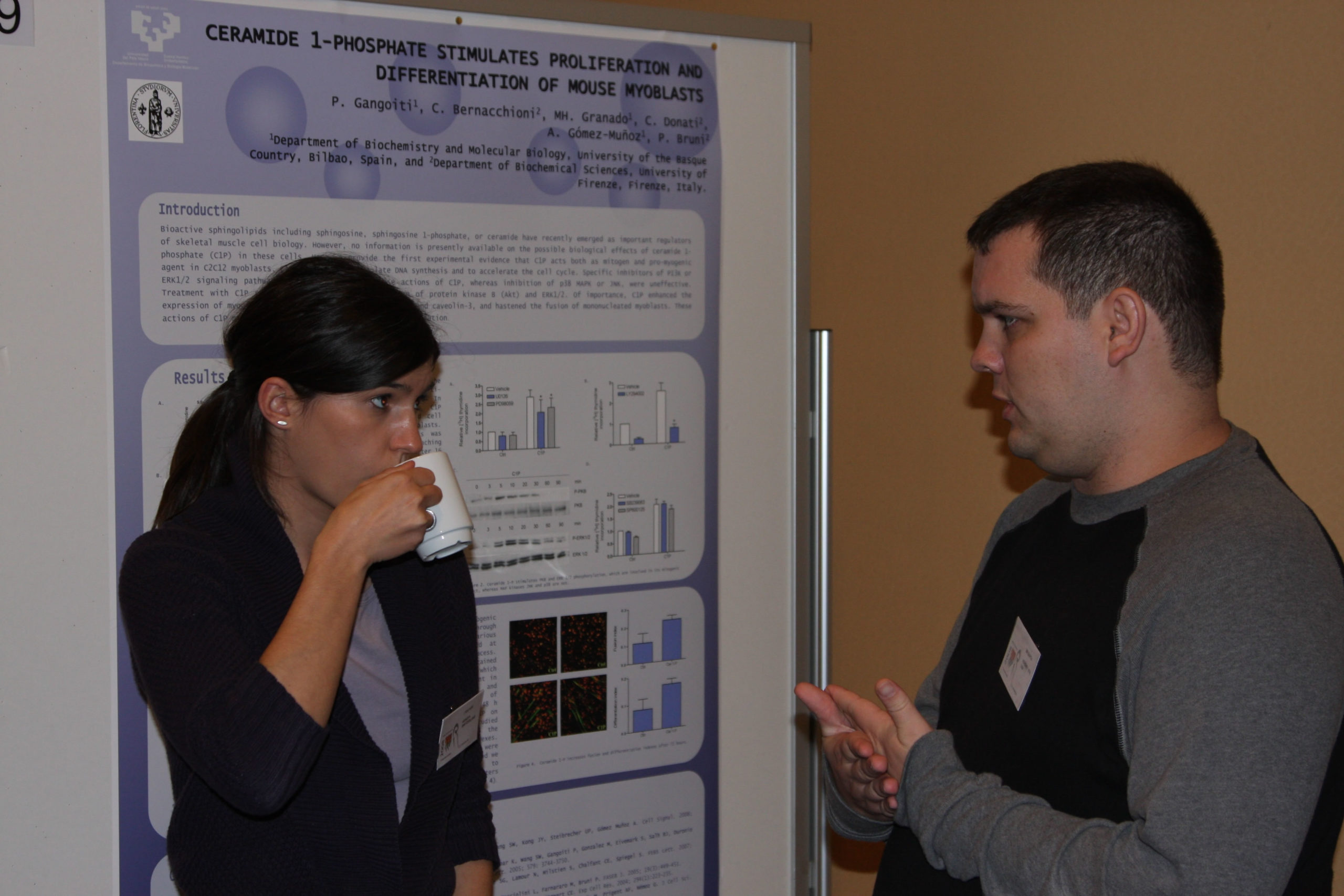
Ceramide 1-phosphate stimulates proliferation and differentiation of mouse myoblasts.
P9
P Gangoiti1, C Bernacchioni2, MH Granado1, C Donati2, AGómez-Muñoz1, P Bruni2
1Department of Biochemistry and Molecular Biology, University of the Basque Country, Bilbao, Spain and 2Department of Biochemical Sciences, University of Firenze, Firenze, Italy
Bioactive sphingolipids including sphingosine, sphingosine 1- phosphate, ceramide, GM3 ganglioside have been recently emerged as important regulators of skeletal muscle cell biology being capable of regulating key cellular parameters including myoblast proliferation and myogenesis. However, no information is presently available on the possible biological effects of ceramide1-phosphate (C1P) in these cells. Here we provide the first experimental evidence that C1P acts both as mitogen and pro- myogenic agent in C2C12 myoblasts. C1P (15µM) was found to stimulate DNA synthesis and accelerate cell cycle. Specific inhibitors of PI3K or ERK1/2 signaling pathways strongly attenuated the C1P action, whereas pertussis toxin treatment, or inhibition of p38 MAPK or JNK were uneffective. In agreement, C1P was responsible for a rapid phosphorylation of Akt and ERK1/2. Notably, C1P was disclosed to exert also a robust promyogenic effect. It enhanced the expression of myogenic markers such as myosin heavy chain and caveolin-3 and hastened the fusion of mononucleated myoblasts. Thus, in this study a novel important biological action of C1P in myoblasts has been highlighted, which possibly will be exploited in the future to enhance skeletal muscle regeneration.
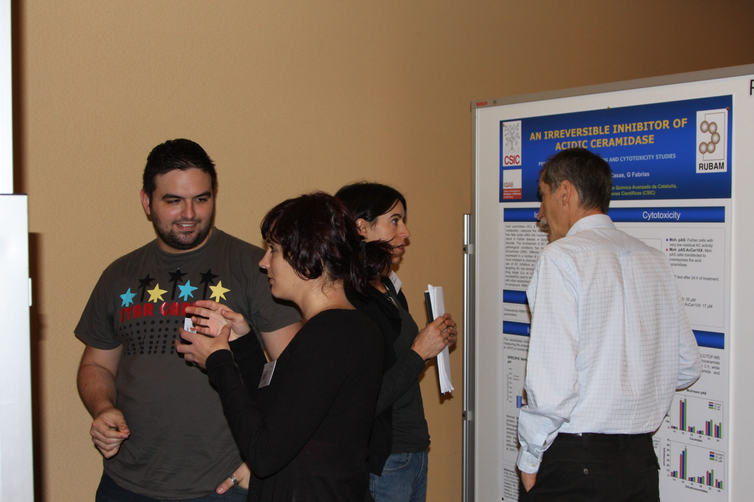
The first irreversible inhibitor of acid ceramidase. Preliminary characterization and cytotoxicity studies.
P10
L Camacho, C Bedia, J Casas, G Fabrias
Dept. of Biomedicinal Chemistry, Instituto de Química Avanzada de Cataluña, Consejo Superior de Investigaciones Científicas (CSIC)
Acid ceramidase (AC) catalyzes the degradation of ceramide into sphingosine and free fatty acids within the lysosomes. Mutations in the AC gene (ASAH1) result in Farber disease, a fatal human genetic disorder. The involvement of AC in cell death in both physiological and pathological conditions has been extensively documented1 and different studies support AC as a drug target in cancer.2 In agreement, AC inhibitors have been successfully used to slow the growth of cancer cells, alone or in combination with other anticancer treatments. Continuing with ongoing projects aimed at the discovery of novel AC inhibitors, in this communication we report on the activity of compound RBM15 as the first irreversible inhibitor of AC. Using Farber cells transduced to overexpress AC, this com- pound inhibited the hydrolysis of CerC12NBD dose- dependently both in intact cells and in cell lysates with IC50 values in the low µM range in both cases.Time-course and dilution experiments proved that inhibition was irreversible. AC inhibition was also evident in the ceramidome, as concluded form HPLC-MS analyses of sphingolipid extracts from cells treated with RBM15. A preliminary account of the cytotoxicity of this molecule over the human adenocarcinoma cell line A459 is presented.
Reduced hiv-1 infection by inhibition of dihydroceramide desaturase.
P11
JM Munoz-Olaya1, C Vieira2, J Sot3, J Casas1, SJimenez-Baranda2, JL Abad1, S Grijalvo1, A Delgado1,4, A Llebaria1, A Alonso3, FM Goñi3, S Mañes2, G Fabrias1
1Dept. of Biomedicinal Chemistry, IQAC-CSIC, Barcelona, Spain – 2Dept. of Immunology and Oncology, CNB-CSIC, Madrid, Spain – 3Dept. of Biochemistry, and Unidad de Biofísica CSIC-UPV/EHU, University of the Basque Country, Bilbao, Spain – 4Pharmaceutical chemistry Unit, School of Pharmacy, University of Barcelona, Barcelona, Spain
HIV-1 infects permissive cells by the gp41-mediated fusion of viral and targets cell membranes. This fusion event requires the sequential interactions of the gp120 viral envelope with CD4, the primary viral receptor, and CXCR4 or CCR5, the coreceptors required for HIV-1 cell entry and infection. Accumulated evidence indicates that lipid rafts have an essential role in the HIV-1-induced lateral associations required for viral infection. Disruption of cell membrane rafts by cholesterol depletion before viral exposure inhibits virus entry. Similar effects are observed with either inhibitors of de novo sphingolipid biosynthesis or inhibitors of glucosylceramide synthase. Here we describe the synthesis of compound GT11pyr and its use as pharmacological tool against HIV cell entry. This compound inhibited the HIV-1 envelope-mediated fusion with target cell membranes in a dose-dependent manner. Biophysical studies with model membranes agree with the biological data. The overall findings reinforce the therapeutical interest of dihydrocer- amide desaturase in anti HIV-1 therapy and other raft-associated diseases.
Acidic and neutral sphingomyelinase inhibition by dihydroceramide and dihydrosphingomyelin analogues.
P12
F Simbari1, X Matabosch1, G Fabriàs1, A Llebaria1, A Delgado1,2, T Levade3, J Casas1
1Dept. of Biomedicinal Chemistry, Instituto de Química Avanzada de Cataluña (CSIC), Barcelona, Spain – 2Unidad de Química Farmacéutica (Unidad Asociada al CSIC), Facultad de Farmacia, Universidad de Barcelona, Barcelona, Spain – 3INSERM U858, Institut Louis Bugnard, Centre Hospitalier Universitaire de Rangueil, Toulouse, France
Sphingomyelin (SM) is the primary sphingolipid in mamma- lian cells and plays important structural and functional roles. SM hydrolysis can be carried out by different isoforms of sphingomye- linase (SMase), producing phosphorylcholine and the intracellular effector ceramide. Ceramide diffuses within membranes acting as a second messenger. Generation of ceramide in various cellular systems is currently recognized as critical to the initiation of vital cellular processes such as differentiation, cell proliferation and apoptosis. In this communication we describe the inhibitory activity of acidic and neutral SMases by dihydroceramide and dihydrosphin- gomyelin analogues, using C6-NBD-sphingomyelin as substrate. Acidic SMase activity was determined with A549 cell homoge- nates whereas homogenates from acidic SMase-deficientNiemann-Pick cells were used as neutral SMase enzymatic source. The most active compounds were tested in intact cells and their effect on the cellular sphingolipid profile was also studied.
Regulation of DHS1P release in F9 mutant cells by fumonisin- B1.
P13
SM Pak1, NY Park1, SM Kim1, KO Shin1, JY Kwak1, HS Yoo1, S Oh2, WJ Kim3, YM Lee1
1College of Pharmacy and CBITRC, Chungbuk National University – 2Department of Neuroscience, College of Medicine, Ehwa Womans University 3Department of Life Sciences, Soonchun- hyang University, Korea
The main goal of our study was to determine the regulation mechanism of dihydrosphingosine 1-phosphate (DHS1P) release from F9 mutant cells. We used a specific mutant (F9-12) of F9 cells over-expressing Sphk1 and showing null of S1P lyase. FB1 treatment (50 uM) exhibited concurrent increases of DHS and DHS1P in F9-12 cells. In this condition, the increased DHS and DHS1P also efficiently released outside. ISP-1 and DMS treatment largely attenuated the accumulation of DHS and DHS1P in both inside and outside of cells. FB1 treatment induced a small percentage of apoptotic cells, indicating the releases of DHS and DHS1P were not mediated by apoptosis. DHS1P release may be regulated by the rate of sphingolipid biosynthesis and Sphk activity. We further investigated the effects of ABC transporters on DHS1P release. By using ABC transporters inhibitors, verap- amil, MK-571 and glyburide, the involvement of ABC trans- porters on DHS1P release was verified.
“This work was supported by the Korea Research Foundation Grant funded by the Korean Government (MOEHRD)” (The Regional Research Universities Program/Chungbuk BITResearch-Oriented University Consortium)
Tumour biology of melanoma: a novel role of acid sphingomyelinase
P14
L Bizzozero1, G Milani2, E Clementi1,3, C Perrotta1
1Department of Preclinical Sciences, University of Milano, Milano, Italy; 2H. San Raffaele Scientific Institute, Milano, Italy; 3E. Medea Scientific Institute, Bosisio Parini, Italy
Recent evidence indicates that Acid Sphingomyelinase (A-SMase),plays important role in tumour biology, including tumour response to chemotherapeutic drugs. The effects of A-SMase were shown in vivo to results from a combination of effects of the enzyme expressed by the tumour cells themselves, and the surrounding microvascular endothelial and inflammatory cells. We decided to investigate the specific role of A-SMase expressed by melanoma B16 cells which show different tumorigenic and metastatic properties dependent on their melanin content. In order to investigate a possible correlation between A-SMase expression and B16 cells phenotype we isolated several B16 clones on the basis of pigmentation, indicating the pigmented clones as “black”, and the not- pigmented as “white” and we observed a higher expression and activity of A-SMase in the white B16 clones. In vivo preliminary experiments showed that the white and the black clones differ in terms of growth rate, invasiveness and histological characteristics. These data suggest a strong relationship betweenA-SMase expression and tumour behav- iour. These studies might help refine therapeutic strategies against tumour based on regulation of A-SMase activity/ expression.
Ceramide induces autophagic death and HMGB1 release in human glioma cells.
P15
P Giussani, R Bassi, V Anelli, F De Zen, L Brioschi, P Viani, L Riboni
Dept Medical Chemistry, Biochemistry and Biotechnology,LITA-Segrate, University of Milan, Milan, Italy
Gliomas are among the deadliest of human cancers, and exhibit striking resistance to therapy. Glioma cells are frequently resistant to apoptosis, the induction of autophagy recently emerging as a potential effective therapeutic approach. The molecular mechanisms underlying autophagic death in glioma cells remain largerly unknown. The aim of this research was to investigate whether the sphingoid mediator ceramide (Cer) may play a role in glioma cell autophagic death. Treatment of human glioma cells with the alkylating agent temozolomide (TMZ) results in the increase of Cer levels, followed by cell death. The administration of short-chainCer analogues mimics TMZ in inducing glioma cell toxicity. After TMZ and Cer treatment, no formation of apoptotic small bodies occurs. Acridine orange staining and western blot of the autophago- some marker LC3B-II indicate TMZ- and Cer- induce autoph- agy. In agreement, co-treatment of cells with the autophagic inhibitor 3-methyladenine reverses toxicity. Moreover, glioma cells undergoing Cer-induced autophagy release large amounts of HMGB1 (high mobility group box 1), after its translocation from the nucleus to cytosol. On its turn, HMGB1 acts as an autocrine stimulus via RAGE receptors, by inducing cell growth and migration. In conclusion, our data implicate Cer as mediator of TMZ-induced autophagic death in glioma cells and suggest glioma cells dying by autophagy provide HMGB1 as extracellular signal to enhance the growth of viable cells.
Desensitization and internalization of sphingosine-1-phosphate receptors induced by cis-4-methylsphingosine.
P16
M ter Braak1, B Hegen1, V Hardel1, KH Jakobs1, G van Echten- Deckert2, D Meyer zu Heringdorf 1,3
1Institut für Pharmakologie, Universitätsklinikum Essen, 2Kekulé- Institut, LIMES Membrane Biology and Lipid Biochemistry, Universität Bonn and 3Institut für Pharmakologie, Universitätsklinikum Frankfurt, Germany
Cis-4-methylsphingosine (cis-4 M-Sph) is a synthetic sphingosine analogue that is readily taken up by cells and phosphorylated to cis-4-methylsphingosine-1-phosphate (cis-4 M-S1P), which accumu- lates intracellularly. Previous reports demonstrated that cis-4 M-Sphmimicked the mitogenic effect of sphingosine-1-phosphate (S1P) in Swiss 3 T3 fibroblasts, but induced apoptosis in neuronal cells. Here, we analysed the influence of cis-4 M-Sph on S1P receptor signalling in HEK-293 cells and mouse embryonic fibroblasts. Addition of cis-4M-Sph to the cells induced increases in intracellular Ca2+concentrations ([Ca2+]i) that were slower than those by S1P but faster than those by sphingosine. Pre-incubation with cis-4 M-Sph, but not with sphingosine, reduced ([Ca2+]i increases by S1P in a time- andconcentration-dependent manner, while it had no influence on ([Ca2+]i increases by LPA. Both the stimulation of acute ([Ca2+]i increases and the inhibition of S1P-induced ([Ca2+]i increases were dependent on the presence of sphingosine kinase-1, indicating that they were mediated by cis-4 M-S1P. Confocal microscopy revealed that cis-4 M-Sphinduced an internalization of S1P receptors. It is concluded that cellular production of cis-4 M-S1P can lead to S1P receptor desensitization and internalization.
Space environment and lipid domains rich in sphingomyelin-cholesterol content.
P17
E Albi1, M Peverini1, R Lazzarini1, M Viola Magni1, G Perrella2
1Department of Clinical and Experimental Medicine, Physiopathology Section, University of Perugia. – 2Department of Experimental and Clinical Pathology and Medicine, University of Udine
It has been shown that in FRTL-5 cells UV treatment induced an increase ofN-SMase activity higher in proliferating cells (TSH+) than in quiescent cells(TSH-). Differently, in the space the enzyme activity increase of TSH+ cells was similar to that of TSH- cells. The aim of the present work was to evaluate the effect of the cosmic environment on lipid domains in relation to FRTL-5 TSH+cell activity during the 15-days on the Space Shuttle (Experia Mission).The activity of N-SMase and the content of RNA polymerase II were evaluated in the cells whereas the lipid composition was analyzed in the culture medium at the re-entry of the shuttle or after 48 hours of cell culture at 37 C. The results showed low value of N-SMase activity and RNA polymerase II content. After 48 hours at 37 C the SMase activity and the content of RNA polymerase II increased in the cell. In the culture medium the content of PE, PI, and PS was similar in the two experimental conditions whereas the PC, SM and CHO content decreased in the space with respect to the medium of the cells after 48 hours at 37 C. It is possible that in space the cells are hungry of lipid domains that are able to make them more stable, slowing down the proliferative activity and protecting them from the damage.
Role of the third amino acid of the e/dry motif in g protein selectivity of s1p receptors.
P19
M Jongsma, FS den Boon, PB van Loenen, MC Michel, SLM Peters, AE Alewijnse
Dept. Pharmacology & Pharmacotherapy, Academic Medical Center Amsterdam, The Netherlands
Sphingosine-1-phosphate (S1P) receptors show variance in the third amino acid of the conserved E/DRY motif. This study investigates whether this amino acid is important in G-proteinselectivity of these receptors. The most remarkable differences were observed for the S1P1 mutant receptors. The S1P1ERH and S1P1ERA mutant receptor showed a decrease in the potency of S1P and SEW2871 to inhibitforskolin-induced cAMP accumulation whereas the potency for FTY720-P was unchanged. The potency of S1P and FTY720-P to induce increases in intracellular calcium concentrations were shifted to the left at the S1P1ERH mutant receptor. The ERH to ERY or ERA mutations of the S1P2 and S1P3 receptor did not induce major changes in signaling. In conclusion, this study is a first indication that the tyrosine residue of the E/DRY motif may restrict the S1P1 receptor from activating multiple G-proteins. However, introducing a tyrosine residue in the S1P2 or S1P3 receptor did affect but did not limit the signaling of these receptors. Interestingly, besides these findings we show that some mutations differentially affected the signaling of the tested ligands.
Importance of connexin-43 in the regulation of sphingolipid metabolism in primary skeletal muscle cells.
P20
F Bini1, J von Maltzahn2, V Wulf2, K Willecke2, Meacci E1
1Department of Biochemical Sciences, University of Florence, Italy; 2Institut für Genetik, Abteilung Molekulargenetik, Universität Bonn, Germany; elisabetta.meacci@unifi.it
Diverse intercellular signalling pathways influence the regulation of myoblast differentiation of skeletal muscle during development and regeneration. One of them is mediated by gap junctions, i.e. intercellular conduits formed by docking of two hemichannels composed of connexin (Cx) proteins. The expression of Cx43 is upregulated during the regeneration of adult skeletal muscle. In vitro studies revealed that Cx43 expression is required for normal myo- genesis. In agreement with this evidence, we have recently shown that the regulation and assembly of connexins into gap junctions and Cx43 protein in particular represent critical events in myoblast differentiation elicited by sphingosine 1-phosphate (S1P), a bioactive lipid involved in this biological process and in the activation of muscle stem cells. Here we investigated the possible interaction between Cx43 expression and function on sphingolipid metabolism in primary cultures of myoblasts obtained from wild type andCx43-knockout (Cx43-/-) newborn mice. Interestingly, we found that sphingolipid metabolism is significantly altered in Cx43-/- cells compared to wild type and heterozygous Cx43-/+ cells. These findings suggest that the ability of myoblasts to share information and/or signalling pathways mediated by Cx43 is required for the correct balance between sphingolipids, the ceramide and S1P. Ceramide and S1P act in opposite manner regulating the coordinated differentiation of myoblasts to their final fate.
(Grants from Fondazione Cassa di Risparmio di Pistoia e Pescia, Programma Vigoni)
S1P and its analogs are killing neurons.
P21
N Hagen, AH Merrill Jr1, G van Echten-Deckert
Kekulé-Institute,LIMES Membrane Biology & Lipid Biochemistry, University Bonn, Germany
1School of Biology and Petit Institute for Bioengineering and Bioscience, Georgia Institute of Technology, Atlanta, USA
Sphingosine-1-phosphate (S1P) is known to induce proliferation in most cell types described so far. However, in the present study we demonstrate that terminally differentiated neurons, which store S1P as a result of S1P-lyase deficiency, are directed into apoptosis. Using liquid chromatography (LC) electrospray ionization (ESI) mass spectrometry we determined the amounts of different bioactive sphingolipids in neurons prepared from lyase deficient versus wildtype and heterozygous mice. Our results confirm the hypothesis based on results obtained with the metabolically stable sphingoid phosphate cis-4-methylsphingosine-phosphate that S1P induces neuronal apoptosis when it reaches a certain threshold value. In addition the molecular mechanism responsible for this effect involves aberrant cell cycle activation as described before in wild type neurons accumulating the semi-synthetic cis-4-methylsphingo-sine-phosphate. Of interest, FTY720 showed a similar yet less pronounced apoptotic effect in wild type neurons. We thus propose cis-4-methylsphingosine as a new synthetic prodrug, which might help understand the role of S1P in neuronal cell death.