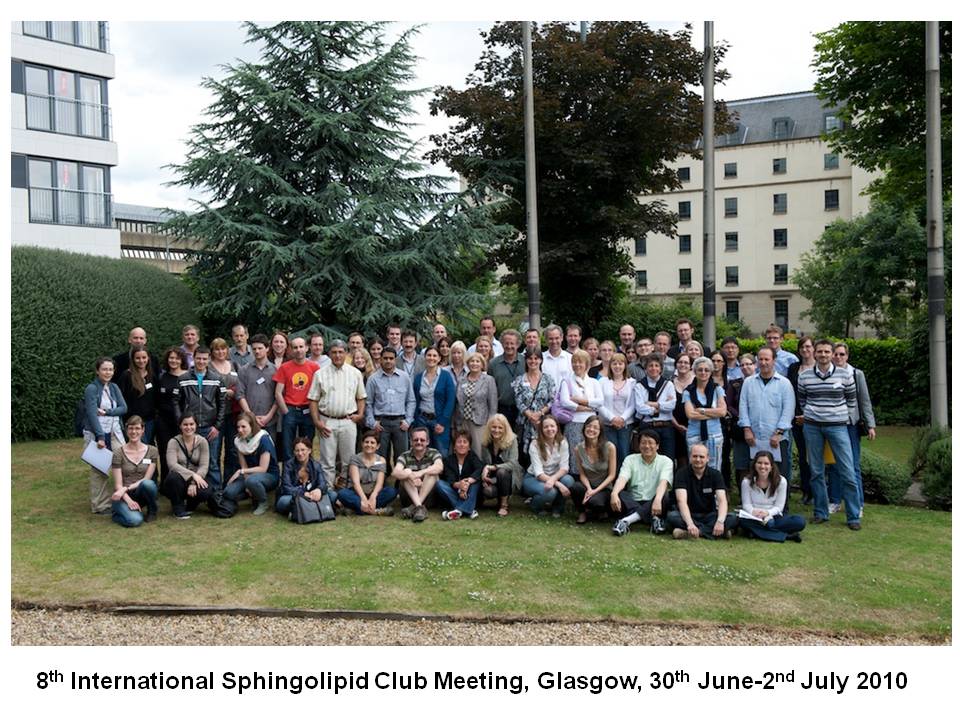
8th Meeting
Glasgow, Great Britain, Nov 14-16, 2010
Partecipants:
1.Elisabeth Schmitz 2. Lide Arana 3. Noemi Jimenez 4. Ilaria Piano 5. Gemma Fabrias 6. Karoly Liliom 7. Paola Bruni 8. Gerhild van Echten-Deckert 9. Anna Pshezhetskiy 10. Francesca Tonelli 11. Yong-Moon Le 12. Dimitry Pshezhtskiy 13. Laura Martin 14. Betul Kavun Ozbayraktar 15. Alberto Ouro16. Joao Nunes 17. Hervé Le Stunff 18. José Luis Abad 19. Lingaraju Marlingapla Halasiddappa 20. Chiara Donati 21. Alice Aòessenko 22. Susan Pyne 23. Giuseppina Perella 24. Carolyn Loveridge 25. Elisabetta Meacci 26. Mercedes Garcia Gil 27. José Fernandez-Checa 28. Nicolas Loiseau 29. Laura Bizzozer 30. Denise Cazzato 31. Jenny Flygare 32. Lysann Saue 33. Julien Veret 34. Christine Wirrig 35. Carmen Bedia 36. Antonio Gomez-Muñoz 37. Sonia Hernandez 38. Elita Avota 39. Astrid Alewijnse 40. Nigel Pyne 41. Erich Gulbins 42. Marya Shupik 43. Keng Gat Lim 44. Carole Mooney 45. Manola Peverini 46. Kati Kemppainen 47. Stephanie Schwalm 48. Dagmar Mayer zu Heringdorf 49. François Paris 50. Riccardo Ghidoni 51. Thierry Levade 52. Guillermo Velasco 53. Keith Mascall 54. Graeme Nixon 55. Gerald Dubois 56. Stephan Peters 57. Michael Jakobi 58. Anja Lüth 59. Iris Fischer 60. Burkhard Kleuser 61. Kid Törnquist 62. Christoph Arenz 63. Nina Bergelin 64. Seikwan Oh 65. Frederik Claas
Attending but not on photo:
1. Stephan Ladish 2. Masa Tada 3. Sandrine Pizette
Regulation of pulmonary inflammation in cystic fibrosis by ceramide.
I1
Erich Gulbins, Yang Zhang, Katrin Anne Becker
Dept. of Molecular Biology, University of Duisburg-Essen, Hufelandstrasse 55, 45122 Essen, Germany
Cystic fibrosis (CF), caused by mutations of the CF transmembrane conductance regulator (CFTR) molecule, is characterized by chronic pulmonary inflammation, reduced mucociliary clearance, and increased susceptibility to infection. We have recently demonstrated an important role of sphingolipids in CF. Cftr-knock-out or B6.129P2(CF/3)-CftrTgH(neoim)Hgu mice that produce low levels of Cftr accumulate ceramide in their lungs. This ceramide triggers enhanced cell death of respiratory epithelial cells, release of pro-inflammatory cytokines in the lung, release of DNA into the mucus, chronic inflammation and high sensitivity to P. aeruginosa infections. Human post-mortem specimens also have high levels of ceramide in lung tissue of CF patients. Using mass spectrometry, fluorescence microscopy and biochemical measurements, we show an accumulation of ceramide also in alveolar macrophages, the trachea and intestinum of CF mice. Ceramide induces a constitutive activation of caspase 1 and an increased rate of cell death in CF mice, which might result in inflammation and increased susceptibility of CF mice. Heterozygosity of the acid sphingomyelinase or systemic application or inhalation of inhibitors of the acid sphingomyelinase reduced ceramide levels in bronchial epithelial cells of CF mice. Normalization of ceramide levels prevented inflammation in the lung of CF mice and infection with P. aeruginosa. These studies indicate a central role of ceramide in the pathophysiology of CF and suggest the acid sphingomyelinase as a novel target to treat cystic fibrosis.
Glycosylation and signaling: glycosphingolipids get their share.
I2
Sandrine Pizette1
1Institute of Developmental Biology and Cancer, Universite de Nice Sophia Antipolis, France
Studies of the Notch receptor have shown that glycosylation of the receptor or its ligand is a means to regulate ligand-receptor interaction. To address whether glycosylation is a general mechanism controling signaling, we undertook the characterisation of two Drosophila genes, egghead (egh) and brainiac (brn), which harbor sequence similarities to genes encoding glycosyltransferases. In addition, mutations in these genes result in phenotypes reminiscent of alterations in Notch and EGFR signaling.
Using in vitro enzymatic assays, we first demonstrated that Egh and Brn are glycosyltransferases that catalyze the synthesis of disaccharides specific to the glycosphingolipid (GSL) pathway. We then showed by biochemical analysis of GSL species and by immunofluorescence with an antibody specific to a short form of GSL, that the egh and brn mutant animals accumulate truncated GSLs. This indicates that Egh and Brn act on GSL biosynthesis in vivo. Finally, we made use of the egh and brn mutants to investigate the role of GSLs in signaling in vivo. We found that during oogenesis, GSLs shape the extracellular gradient of the Drosophila TGFa-like EGFR ligand by controling its diffusion. These results thus reveal an unexpected role for GSLs in signaling.
I will discuss the molecular mechanism underlying this novel function, and will present preliminary data indicating new in vivo roles for GSLs at both the organismal and subcellular level.
Ceramide kinase, ceramide-1-phosphate: roles and regulation.
I3
Frederic Bornancin
Novartis Institutes for BioMedical Research, Basel, Switzerland
It has been almost a decade since ceramide kinase (CERK) was cloned. Phosphorylation of ceramide by CERK remains the only known mechanism for production of ceramide 1-phosphate (C1P). There is, however, mounting evidence for an as yet unidentified alternative C1P producing pathway. Therefore, the knowledge acquired using C1P as active principle may only partially reflect modulation of CERK. Nevertheless, CERK appears to be key for controlling ceramide levels, and C1P produced by CERK has emerged as a genuine signaling entity.
The current understanding of CERK at protein level as well as some insights into the regulation of ceramide metabolism by CERK will be presented, including new data on the modulation of CERK in innate immunity.
Tumor gangliosides condition the microenvironment and favor tumor progression.
I4
Stephan Ladisch
Children’s Rresearch Institute, Children’s National Medical Center, 111 Michigan Ave. NW, Washington, DC, 20010, USA
To better understand the pathogenesis of human cancer, increasing attention has been directed to identifying potential in vivo interactions between the tumor cell and the surrounding tumor microenvironment–-the tumor-host interaction. Our findings on the role of key membrane molecules, gangliosides, in influencing these tumor-host interactions will be presented. Briefly, by their synthesis and subsequent shedding from tumor cells, tumor cell gangliosides enhance, and interference with their synthesis impedes, tumor development and progression in vivo. Specifically, the characteristically rapid tumor cell ganglioside metabolism, i.e., substantial synthesis and shedding into the tumor microenvironment, results in transfer to surrounding normal cells. Subsequently, these ganglioside-enriched normal cells have altered functions. Basic cellular mechanisms of ganglioside effects in the tumor microenvironment–-inhibition of the immune response, enhancement of stromal cell proliferation, and enhancement of the angiogenic response–-will be highlighted. The critical question raised is: Is tumor formation and progression itself affected? To directly address this, we created a genetically stable, complete, and specific model of tumor cell ganglioside depletion. Early findings on the consequences of selective and complete depletion of tumor cell gangliosides in this murine sarcoma cell model will be summarized, and their implications for cancer treatment considered. [NIH grants R01CA42361-17 and R01 CA61010-14.]
Targeting the sphingosine 1-phosphate signalling pathway in cancer.
I5
Nigel J. Pyne, JS Long, C. Loveridge, F. Tonelli, KG Lim, S. Pyne
Cell Biology Group, Strathclyde Institute of Pharmacy and Biomedical Sciences, University of Strathclyde, 27 Taylor St, Glasgow, G4 0NR, Scotland, UK.
Sphingosine 1-phosphate is a bioactive lipid that is formed by the sphingosine kinase-catalysed phosphorylation of sphingosine, and which binds to a family of G-protein coupled receptors termed S1P1-5 to elicit biological responses. We will demonstrate in this presentation that high expression of S1P1, S1P3 receptors and sphingosine kinase 1 (SK1) in ER+ breast cancer tumours are associated with reduced patient survival and recurrence on tamoxifen when compared with patients with low expression of these proteins in their tumours. We will also provide evidence for how S1P1 receptors participate in tumour neovascularisation and how SK1/S1P3 function together to induce a metastatic phenotype in ER+ breast cancer cells. A novel promiscuous interaction between S1P4 and the HER2 oncogene in ER- breast cancer cells will also be described. The highlighting of the role of SK1 and S1P receptors in breast cancer provide impetus for targeting these proteins with compounds that inhibit their function. In this regard, we will also demonstrate novel actions of SK1 inhibitors, which reveal a new mechanism by which SK1 induces chemotherapeutic resistance in androgen-independent prostate cancer cells.
Supported by CRUK [A7536].
Dual and opposite effects of fumonisin B1 on ceramide and sphingomyelin contents in piglet’s lung and liver.
S1
N. Loiseau1, N. Therville2, T. Levade2, J. Bertrand-Michel3
1 UR66, Laboratoire de Pharmacologie-Toxicologie, INRA,
2 U858, Institut de Médecine Moléculaire de Rangueil, INSERM,
3 U563, Plateau technique de Lipidomique, IFR 30, INSERM, Toulouse, France
Fumonisin B1 (FB1) is one of the most known inhibitor of Ceramide Synthases (CerS). In this study, we hypothesized that FB1 may interact with a specific CerS subgroup to explain the interspecies differences in the clinical symptoms.
Since each CerS uses a preferential subset of fatty acyl-CoAs, we compared the sphingolipid contents of lungs and liver from normal and FB1-contaminated piglets (fed with 1.5 mg FB1/kg body weight daily for 9 days). This study focused on the analysis of type 2 ceramide (Cer2) and sphingomyelin (SM) contents. Total Cer2 and total SM contents decreased 2-fold and 1.6-fold, respectively in lungs of FB1-contaminated piglets. In contrast, surprisingly, in liver of FB1-contaminated animals, total Cer2 and total SM contents increased 5.8-fold and 1.5-fold, respectively. By analyzing the FB1 effects on individual Cer2 and SM species, we deduced the effect of this toxin on each CerS family. Our data indicate that FB1 inhibits CerS5/6 whatever the tissues. Moreover, this toxin interacts with piglet’s CerS2/4 to deplete lung and to enrich liver with ceramides containing long fatty acids. These changes could explain the species-specific toxicity of FB1.
[Supported by DAER-Recherche/06001316/07006301]
Increased expression of sphingosine kinase 1 mediates prosaposin-induced cytokine production.
S2
Lysann Sauer1, Dmitry Pshezhetskiy1 and Jonathan Waxman1
1 Tumour Microenvironment and Chemotherapy Group, Hammersmith Campus, Faculty of Medicine, Imperial College London, UK
Sphingosine kinase 1 (Sphk1) upregulation has been implicated in chemo-resistance in prostate cancer and its inhibition can sensitize to chemo-induced apoptosis. Prosaposin, a neurotrophic protein has been recently shown to induce prostate cancer cell proliferation, migration and chemoresistance. Furthermore an increased expression of prosaposin was linked with progression of human prostate cancer.
Prosaposin knockout mice exhibited an involution of the prostate gland which correlated with a significant reduction of prostate SphK1 activity. In prostate cancer cell lines SphK1 activity correlated with the amount of secreted, but not intracellular prosaposin, indicating a role for the prosaposin/GPCR signalling. The specific knockdown of prosaposin in prostate cancer cells induced a significant decrease in SphK1 activity and expression on mRNA and protein level. Additionally, prosaptide TX14A, derived from the trophic sequence of saposin C, enhanced SphK1 activity and up-regulated SphK1 mRNA expression via ERK1/2-mediated mechanism. Furthermore, TX14A induced an up-regulation of the proliferative factor interleukin 6, which was abrogated by pre-treatment with SphK1-specific siRNA.
Overall, our data indicate that SphK1 upregulation is required for prosaposin-induced cytokine expression in prostate cancer cells.
Role of sphingolipid enzymes in oxidized phospholipid (oxPL) induced RAW 267.4 macrophage cell death.
S3
L Marlingapla Halasiddappa,D Koller, U Stemmer, Z Dunai, E Zenzmaier, A Hermetter
Institute of Biochemistry, Graz University of Technology, Petersgasse 12/2, 8010 Graz, Austria.
Oxidized lipoproteins and their oxidized phospholipids (oxPL) induce apoptosis in vascular cells. Ceramide (Cer) mediates and propagates apoptosis by activation of JNK and p38 MAPkinases. Cer is generated instantly by aSMase via sphingomyelin hydrolysis and by de novo synthesis in the hours. Ceramide synthase (CerS) isoforms regulate de novo generation of ceramides as they display substrate specificity for the chain lengths of fatty acyl-CoAs. Depending on the stimulus and cell type, specific ceramide species synthesized by these isoforms (CerS1-6) are likely to contribute differently to the apoptosis pathway. Here we report that only a subset of CerS is activated in RAW 264.7 cells by oxPL treatment in a time-dependent manner. Levels of apoptotic ceramide are also influenced by modification and/or degradation. So far, we have studied the activities of ceramidase in oxPL-treated macrophages. This enzyme is almost unaffected and seems not to play any particular role in ceramide-induced cell death. Currently we study the role of other enzymes regulating the apoptotic ceramide pool in cultured RAW 267.4 macrophage cells under the influence of oxPL (POVPC and PGPC). Analyzing the sphingolipidome, as well as expression, localization and activity at various stages of programmed cell death should help understanding the toxicity of oxPL and role of sphingolipid species in mediating apoptosis.
Involvement of SphK1 in LPS-induced TLR 4-mediated accumulation of HIF-1α protein, activation of ASK1 and production of IL-6.
S4
Pshezhetskiy D1*, Nunes J1, Coughlan K2, Lall H2, Waxman J1, Sumbayev V2*
1 Dept Oncology, ICL,
2 Medway School of Pharmacy, Univ. Kent
Toll-like receptors (TLRs) lie at the core of resistance to infectious diseases detecting various pathogens. While both plasma membrane associated TLR4 (recognises bacterial LPS) and endosomal TLR7/8 (recognise viral single-stranded RNA) induce expression of proinflammatory cytokines through redox-dependent upregulation of HIF-1α, the intracellular mechanisms mediating this expression vary. Recently sphingosine kinase 1 (SphK1) was reported to act downstream of TLR4.
In this present study we have identified the implication of the SphK1 signalling in TLR-mediated inflammatory response. In THP-1 and RAW264.7 macrophages ligand-induced activation of the TLR4 but not TLR7/8 induced activation and transcriptional upregulation of SphK1. Activation of SphK1 was dependent on both ERK and PLC-1γ/PI3 kinase pathways and in turn mediated production of ROS and an increase in HIF-1α expression and ASK1 activity. Importantly, TLR4-mediated SphK1 activation was critical for the prevention of LPS-induced activation of caspase 3 and the expression of pro-inflammatory cytokine IL-6.
In conclusion, our findings suggest a novel SphK1-mediated mechanism of TLR4-induced activation of ASK1/HIF-1α axis and cytokine production. Our data strongly suggest that SphK1 inhibition may prove effective to fight bacterial sepsis.
Involvement of the S1P3 receptor in neuroinflammation.
S5
I Fischer1, C Alliod1, N Martinier1, M Frossard1, C Brana1, S Pouly1
1 TA Neurodegenerative Diseases, Merckserono International S.A., Switzerland
Reactive astrocytes are implicated in the development and maintenance of neuro-inflammation in multiple sclerosis (MS) lesions. The sphingosine signalling pathway, including the S1P3 receptor was shown to be involved in the mediation of the inflammatory response in different cell types. So far, the potential role of S1P3 receptor signalling in reactive astrocytes has, however not been defined.
Our data show for the first time the specific expression of the receptor S1P3 on astrocytes in human MS-brain tissue. In cultures of primary rat astrocytes, the treatments with the pro-inflammatory stimuli LPS or with a combination of TNFα and IFNγ increased the mRNA expression of SphK1 and S1P3 receptor. In addition, the protein expression of the SphK1 and S1P3 were increased on the plasma membrane, suggesting increased signalling. Furthermore, LPS treatment increased specific ERK1/2-phosphorylation, which could be mediated via S1P3 as demonstrated by using a S1P3 specific agonist. Moreover LPS, cytokines or sphingosine-1-phosphate (S1P) induced the migration of astrocytes and the release of CXCL1, a known inducer of oligodendrocyte proliferation and chemo-attractant for immune cells.
Together these findings provide new evidence for the implication of SphK1 and S1P3 signalling in reactive astrocytes under inflammatory conditions.
Sphingosine 1-phosphate induces differentiation of mesoangioblasts towards smooth muscle cells.
S6
Donati C1,2., Marseglia G.3, Magi A.3, Cencetti F.1,2, Bernacchioni C.1,2, Benelli M.3, Torricelli F.3, Bruni P.1,2
1 Dip. Scienze Biochimiche, 2Istituto Interuniversitario Miologia (IIM), Università di Firenze and 3UO Citogenetica e Genetica AOUC Firenze, Italy
Smooth muscle cells (SMCs) control fundamental functions such as arterial tone and airway resistance. Recent studies proved that circulating, SMC progenitor cells can contribute to tissue repair following vascular injury. Mesoangioblasts (Mbs) are a new type of stem cells, capable of differentiating into mesoderm cell types, such as muscle and bone. Sphingosine 1-phosphate (S1P) is a lipid mediator that regulates many biological processes as vascular development and SMC growth and migration. We previously demonstrated that S1P acts as potent mitogen and antiapoptotic agent in Mbs. We also showed that TGFβ exerts a marked antiapoptotic action in Mbs, involving the regulation of SK1. In order to exploit the therapeutic potential of these cells, we performed a microarray study to establish transcriptional profiles of human Mbs treated with S1P for 6 h and 24 h. Obtained result, validated by Real Time PCR, Western blotting and immunofluorescence analysis, demonstrate that S1P promotes differentiation of human Mbs towards SMC. Moreover, we provide here evidence that TGFβ-induced differentiation of Mbs into SMC relies on SK regulation. This study highlights a new role for S1P in Mbs which can be used to favour vascular regeneration.
Pharmacological regulation of sphingolipid metabolites in blood.
S7
YM Lee1,T Hla2, K Venkataramann2, S Oh3, SM Kim1, KO Shin 1, HS Yoo1.
1 College of Pharmacy and CBITRC, Chungbuk National Univ.,
2 Center for Vascular Biology, Cornell Univ.,
3 Dept. Neurosci. and Medical Research Institute, Ewha Womans Univ., South Korea
Intracellular and extracellular S1P is tightly regulated by the relative enzyme activities of Sphk1 and Sphk2, S1P lyase and lipid phosphate phosphohydrolases. Extracellular S1P exported by the aid of ABC transporters binds to S1P receptors to transfer its signaling. Recent studies have concluded that S1P gradient and S1P receptor signaling enable immune cell egress from lymphoid tissues. However, the maintenance and regulating factors on S1P level in blood is still under investigation. We found that anti-Fas antibody (Jo2) or combination of LPS and D-GalN treatment into C57/BL6 mice significantly induced liver damage and showed reduced plasma S1P level, but not plasma ceramides. The forced exercise also reduced sphingolipid metabolites including S1P in plasma. The combined treatment of LPS and D-GalN greatly reduced Sphk1 activity in whole blood, while no changes were observed in plasma Sphk1 activity. Notably, the half-life of injected C17-S1P in this condition was not changed. The Sphk1 activities in RBCs and cultured HUVEC cells were not significantly changed by Jo2 or LPS+D-GalN treatment. The Sphk1 activity in liver tissues was slightly increased after liver injury. However, partial hepatectomy did not reduce plasma S1P concentration. Thus, Sphk1 activity in whole blood mainly participated to maintain plasma S1P level. In future the pharmacological effects of ABC transporters inhibitors and Sphk2 activity will be investigated.
Sphingosine 1-phosphate inhibits angiogenesis via interaction with vascular smooth muscle cells.
S8
KS Mascall, GR Small, GF Nixon
Institute of Medical Sciences, University of Aberdeen, Aberdeen, UK
Following myocardial infarction, new blood vessels sprout from coronary arteries. Sphingolipids released from the blood clot could have an effect on this response. Evidence shows that sphingosine 1-phosphate (S1P) may be an important pro-angiogenic factor. This is predominantly through the actions of S1P1 receptors although the S1P2 receptor subtype may inhibit angiogenesis. The aim of this study was to examine the effects of S1P on angiogenesis using ex vivo human arteries and a co-culture in vitro model.
In human mammary artery placed in Matrigel, S1P inhibited endothelial tubule formation. Co-culture of primary cultured human fibroblasts, human coronary artery smooth muscle (HCASM) cells and human coronary artery endothelial cells also revealed a significant inhibition of endothelial tubule formation following treatment with S1P. When HCASM cells were omitted from the co-culture, tubule formation was not affected by S1P incubation. Incubation with specific antagonists determined that this inhibition occurred through activation of S1P2 receptors linked to the rho-kinase pathway. Conditioned medium from S1P-treated HCASM cells added to the co-culture also inhibited endothelial tubule growth suggesting the inhibitory factor(s) are released from smooth muscle cells. In conclusion, S1P inhibits angiogenesis through activation of S1P2 receptor and this involves interaction with smooth muscle cells. Such sphingolipid-mediated inhibition of angiogenesis may occur following myocardial infarction.
Sphingosylphosphorylcholine, but not sphingosine 1-phosphate, is a pro- inflammatory mediator in rat cerebral artery vascular smooth muscle.
S9
C Wirrig, FA Mathieson, I Hunter, GF Nixon
Institute of Medical Sciences, University of Aberdeen, Aberdeen, UK.
Following subarachnoid haemorrhage (SAH) of the cerebral arteries a marked inflammation occurs. This is caused by an adventitial blood clot and contributes to the subsequent cerebral artery vasospasm and ischaemia. Pro-inflammatory mediators in SAH have not yet been identified. As sphingosylphosphorylcholine (SPC) and sphingosine 1-phosphate (S1P) are elevated in serum, we investigated the pro-inflammatory potential of these sphingolipids in rat cerebral arteries using an ex vivo model.
SPC, but not S1P, induced activation of p38 mitogen-activated protein kinase (MAPK), a kinase involved in inflammatory signalling. A transcription factor array identified SPC-induced DNA binding for the transcription factors nuclear factor-κB and CCAAT-enhancer-binding proteins in cerebral arteries, confirmed by electromobility shift assays. S1P did not increase the activity of these transcription factors. In the rat vascular smooth muscle cell line, A7r5, an inflammatory protein array revealed that SPC elicited release of only one pro-inflammatory chemokine, monocyte chemotactic protein (MCP)-1. Further investigation also showed SPC-induced MCP-1 production in rat cerebral arteries.
In summary we demonstrate that SPC can act as a pro-inflammatory mediator in cerebral arteries via increased activity of inflammatory transcription factors and subsequent release of MCP-1. This may contribute to vasospasm following SAH.
C1P and its synthetic analog, PCERA-1, have distinct receptors in macrophages.
S10
T Zor1, A Gómez-Muñoz2, M. Meijler3, H. Rosen4, D Avni1, A Philosoph1, M Levi1, L Arana2, A Ouro2
1 Dept. Biochemistry, Tel-Aviv University, Tel-Aviv, Israel,
2 Dept. Biochemistry & molecular biology, University of the Basque country, Bilbao, Spain,
3 Dept. Chemistry, Ben-Gurion University, Be’er-Sheva, Israel, and
4 Dept. of Immunology, The Scripps Research Institute, La Jolla, CA, USA
Tight regulation of the production of pro- and anti-inflammatory cytokines is essential for the prevention of chronic inflammatory diseases. A synthetic C1P analog, named PCERA-1 (Phospho-CERamide Analog-1), suppressed production of the pro-inflammatory cytokine TNFα, and elevated production of the anti-inflammatory cytokine IL-10, in LPS-stimulated macrophages. PCERA-1 and bovine brain-derived C1P activated distinct signaling pathways in RAW264.7 macrophages. PCERA-1 modulated cytokine expression via the Gs protein and the cAMP pathway, whereas C1P stimulated macrophages migration via the Gi protein and the NFκB pathway. Neither PCERA-1 nor C1P mimicked or antagonized the activities of each other, and PCERA-1 failed to interfere with a C1P binding assay. These results thus indicate that PCERA-1 and C1P bind and activate distinct GPCRs expressed in RAW264.7 macrophages. The orphan receptor GPR3 is predicted to be a phospholipid-binding receptor, due to sequence homology with the S1P and LPA receptor families. Our preliminary results indicate that GPR3 is a receptor for PCERA-1, and thus suggest that GPR3 regulates inflammation.
Ceramide 1-phosphate stimulates reactive oxygen species (ROS) formation. implication in macrophage growth.
S11
L Arana, A Ouro, P Gangoiti , A Gomez-Muñoz
1Dept. Biochemistry and Molecular Biology, Faculty of science and technology, University of the Basque Country, 48080 Bilbao, Spain
The pro-mitogenic and anti-apoptotic actions of Ceramide 1-phosphate (C1P) are now well established. Major pathways involved in the mitogenic effect of C1P include mitogen-activated protein kinase kinase (MEK)/extracellularly regulated kinases (ERK1-2), phosphatidylinositol 3-kinase (PI3K)/protein kinase B (PKB, also known as Akt), c-Jun N terminal kinase (JNK), and protein kinase C-alpha. We report here that C1P induces ROS formation through activation of NADPH oxidase. C1P-stimulated ROS production was inhibited by the NADPH oxidase inhibitor apocynin, the cell-permeable ROS scavenger N-acetyl cystein (NAC), the protein kinase C (PKC) inhibitor Go6976, the PKC-delta inhibitor rottlerin, and by long-term treatment with the phorbol ester PMA, a condition known to downregulate PKC activity. Moreover, a specific cytosolic phospholipase A2-alpha inhibitor potently blocked C1P-stimulated ROS production, and all of the ROS inhibitors blocked C1P-stimulated macrophage proliferation. These findings suggest that ROS are implicated in the mitogenic effect of C1P in macrophages.
This work was supported by grants BFU2009-13314 from MCINN and S-PE09UN42 from the Basque Government (Spain)
Role of the sphingolipid biostat in apoptosis of pancreatic β cells induced by palmitate.
S12
Hervé Le Stunff1, Julien Veret1, Evgeny V. Berdyshev2, Anastasia Skobeleva2, Viswanathan Natarajan2 and Bernard Portha1
1 Laboratoire de Biologie et Pathologie du Pancréas Endocrine, Unité BFA, CNRS EAC 4413, Université Paris Diderot-7, Paris, France.
2 Department of Medicine, Section of Pulmonary and Critical Care Medicine, University of Chicago, Chicago, IL, USA.
Prolonged exposure to fatty acids with high glucose induced pancreatic β cell apoptosis. This study aimed to ascertain the role of sphingolipid in β cell apoptosis induced by palmitate + high glucose. Lipidomic analyses revealed that palmitate but also 30 mM glucose increased de novo ceramide synthesis. Importantly, thirty mM glucose potentiated palmitate-induced accumulation of dihydro-sphingosine and ceramide levels, especially C18:0, C22:0 and C24:1. Apoptosis induced by palmitate with high glucose was partially blocked by fumonisin-B1, an inhibitor of ceramide synthase. Interestingly, palmitate + high glucose stimulated accumulation of dihydrosphingosine-1-phosphate (DHS1P). N,N-dimethyl-sphingosine, a potent inhibitor of sphingosine kinase, increased by 2-fold palmitate + high glucose-induced caspase activity. Furthermore, SphK1 over-expression in β cells drastically increased DHS1P levels and partially inhibited palmitate-induced caspase activation. Together, these results indicate that regulation of the dynamic balance between ceramide and DHS1P levels, the sphingolipid biostat, by palmitate with high glucose concentrations will play a critical role in the survival of pancreatic β cells during the development of type 2 diabetes.
Sphingosine kinase-1 contributes to the anti-fibrotic effect of PPARγ agonists.
S13
A Völzke1, A Koch1, A Huwiler2, J Pfeilschifter1
1 pharmazentrum frankfurt/ZAFES, Goethe University Frankfurt, Frankfurt am Main, Germany
2 Institute of Pharmacology, University of Bern, Bern, Switzerland
Peroxisome proliferator-activated receptor (PPAR)γ agonists (thiazolidinediones; TZDs) are known to act in an anti-fibrotic manner. Previously, it was shown that sphingosine kinase-1 (SK-1) depletion or its pharmacological inhibition led to accelerated expression of the profibrogenic molecule connective tissue growth factor (CTGF) suggesting a protective role of SK-1 activity in the fibrotic process.
Here, we investigated the effect of TZDs on the transcriptional regulation of SK-1 in vitro (rat mesangial cells) and in vivo (mice glomerula) and evaluated the putative role of SK-1 in the anti-fibrotic effect of TZDs. Stimulation with troglitazone and rosiglitazone led to increased SK-1 activity which was preceded by elevated mRNA and protein expression. This effect is due to enhanced SK-1-promotor activity which contains seven putative PPAR response elements. Furthermore, pre-incubation with the PPARγ antagonist GW-9662 inhibited the stimulating effect of TZDs on SK-1 mRNA expression and activity. Finally, the up-regulation of SK-1 by TZDs was paralleled by decreased CTGF expression, an effect which was abolished using the SK-1-inhibitor SKI II. In summary, TZDs dependent SK-1 activation results in lower CTGF demonstrating an essential role of SK-1 in the anti-fibrotic effect of PPARγ agonists.
Synthesis and biological activity of novel inhibitors of acid sphingomyelinase.
S14
Christoph Arenz
Institut für Chemie, Humboldt Universität zu Berlin, Germany
The acid sphingomyelinase (aSMase) is a key enzyme involved in lipid signalling and an emerging drug target.
Many inhibitors of this enzyme are either unspecific, not active in cell culture or have an indirect way of action. We used the potent inhibitor of aSMase, phosphatidyl-3,5-bisphosphate (PtdIns3,5P2) as a lead for the development of novel aSMase inhibitors with improved features. Our inositol- or carbohydrate-based inhibitors are at least of the same potency as PtdIns3,5P2 and inhibit dexamethasone (Dex)-induced apoptosis in HEK-293 cells. In addition, we have synthesized a class of novel bisphosphonate inhibitors that are the most potent inhibitors of aSMase so far. The IC50 for aSMase is 20 nM and >2000fold lower than for nSMAse. The selectivity versus the remote homologue phsophatase PP1 is at least 200fold. Moreover the most potent bisphosphonate completely suppresses Dex-induced apoptosis in HEK-293 cells and inhibits the formation of PAF-induced pulmonary edema in an ex-vivo model. However, many aspects of the biological activity of these substances need to be clarified. So far, the usability of theses inhibitors for medical and in vivo applications is unclear.
Role of sphingolipid trafficking in cannabinoid-induced autophagy.
S15
S Hernández-Tiedra1 , IJ Salanueva1, M Salazar1, G Fabriàs2, J Casas2, K Hanada3, M Guzmán1, G Velasco1
1 Dept of Biochem and Mol Biol I, Complutense University, Madrid, Spain
2 Research Unit on BioActive Molecules, Dept de Química Biomèdica, (IQAC), Barcelona, Spain
3 Dept of Biochemistry and Cell Biology, National Institute of Infectious Diseases, 1-23-1, Toyama, Shinjuku-ku,
Macroautophagy is a conserved degradative process implicated in the turnover of damage organelles and long-live macromolecules. Under starvation conditions, autophagy promotes cell survival providing cells with nutrients derived from the lysosomal degradation of the autophagosome content. However, under specific situations autophagy can lead to cell death.
Work by our laboratory has unraveled that the mechanisms of cannabinoid antitumoral action relies on a ceramide-dependent stimulation of the stress regulated protein p8 and its downstream target TRB3 which leads to stimulation of autophagy and apoptosis in cancer cells. In this work, we investigated whether modulation of sphingolipid trafficking could play a role in the stimulation of autophagy by cannabinoids.
Our results show that THC but not other autophagy inducers alters the sub-cellular distribution of fluorescent analogous of ceramide and promotes the accumulation of long chain sphingolipids species in the microsomal fraction of U87MG glioma cells. Moreover, we also found that THC administration promotes the translocation to the autophagosomes of the ceramide transporter protein (CERT). Our data suggest that changes in sphingolipids distribution play a pivotal role in the stimulation of autophagy-mediated cell death by cannabinoids.
Nuclear import of sphingomyelinase in space environment.
S16
E. Albi1, M. Peverini1, E. Damaskopoulou1, F. Cingolani1, R. Lazzarini1, F. Curcio2, F.S. Ambesi-Impiombato2, G. Perrella2
1Dept. Clinical and Experimental Medicine, Faculty of Medicine and Surgery, Perugia University, Italy and 2Department of Experimental and Clinical Pathology and Medicine, University of Udine, p.le S.M. della Misericordia, 33100 Udine, Italy
It has been reported that during space missions the thyroid cells cultured in the presence of thyrotropin (TSH) decreased their proliferation rate and both their cell growth and lipid metabolism were similar to those of quiescent cells. A rearrangement of cell membrane lipid rafts with an alteration of TSH-TSH receptor interaction has been suggested (1). To study whether the sphingomyelinase (SMase) could be involved in the modification of the cell function in space environment, the SMase localization was analysed either in vitro or in vivo experiments during space TEXUS-44 and STS-129 mission respectively. FRTL5 thyroid cells in culture and thyroid of mice were analysed after the return to earth. The SMase was localized with indirect immunofluorescence by using anti-SMase antibodies alone or in combination with propidium iodine. The results showed that in the ground SMase is localized either in cell membrane or in the nucleus. After space mission the nuclear concentration of this enzyme increases thus indicating a cytoplasmic-nucleus translocation by influencing signal transduction.
1) E. Albi, F.S. Ambesi-Impiombato, E. Damaskopuolou, M. Peverini, A. Lazzarini, R. Lazzarini and G. Perrella. ESA Publications, 2009
Metabolic reconstruction of in silico yeast sphingolipid metabolism incorporating hydroxylation levels of ceramide.
S17
F. Betul Kavun Ozbayraktar1, Kutlu O. Ulgen1
1 Dept. of Chem. Eng., Bogazici University, 34342 Bebek, Istanbul, Turkey
The hydroxylation level diversifies the identity, structure and location of complex sphingolipids in higher eukaryotes. In order to clarify the significance of hydroxylation on sphingolipids, the first metabolic model of Sacchoramyces cerevisiae sphingolipid metabolism was reconstructed in silico, taking into consideration five levels of hydroxylation derived from dihydroceramide, alpha-dihydroceramide, phytoceramide, alpha-phytoceramide and alpha-alpha-phytoceramide. The reconstructed model, which is validated with experimental findings from literature resources, equipped us with the analysis of complex sphingolipid content of the plasma membrane coupled with DAG and phosphatidic acid biosynthesis and ATP consumption rates of in silico cell. These critical metabolites with utmost importance to cell’s viability are utilized as criteria in studying all single deletion mutants of in silico model to propose novel potential drug targets for inducing apoptosis to be further used in cancer therapy. The computational systems biology tools used for interpreting simulation results in terms of their metabolic consequences, flexibility and robustness are flux balance and variability analysis, minimal cut set calculations and principal component analysis. First and second order hydroxylated phytoceramide derived mature sphingolipid synthesis happen to be the most robust part among the others.
Sphingolipids in retinal degenerations.
S18
I. Piano1, E. Novelli2, G. Sala3, P. Gasco4, C. Gargini1, E. Strettoi5, R. Ghidoni3
Dept. Psychiatry & Neurobiology, Univ. Pisa1; G.B. Bietti F’dn for Ophthalmology, Rome2; DMCO, San Paolo Med. School, Univ. Milan3; Nanovector srl, Turin4; Neuroscience Inst. CNR, Pisa5, Italy
Ceramide is a known pro-apoptotic messenger whose de-novo biosynthesis is associated with cell death initiation. In Retinitis Pigmentosa (RP) photoreceptor death occurs by apoptosis. We investigated the role of ceramide in RP using the rd10 mutant mouse, a model of this disease. Myriocin, an inhibitor of serine palmitoyltransferase (SPT, the rate-limiting enzyme of ceramide biosynthesis) was either injected intraocular or administered daily to rd10 mice as eye drops preparations of Solid Lipid Nanoparticles (SLNs). Control mice were administered with vehicle alone or unloaded lipid particles, respectively. We found that retinal ceramide levels double during the time interval of maximum photoreceptor death in rd10 mice (from P14 to P30). Intraocular Myriocin decreases the number of rd10 pycnotic photoreceptors by approximately 50%. Electroretinogram (ERG) recordings were obtained from animals chronically treated with Myriocin-SLNs. ERG a-waves persist after P30 in treated mice while these responses are virtually extinct in control littermates. Confocal microscopy of retinal sections from ERG recorded animals aged P24 (peak of rod apoptosis) up to P30 showed prolonged survival of photoreceptors in treated animals.
This study is the first in vivo demonstration of the possibility to decrease photoreceptor apoptosis by lowering retinal ceramide levels through non-invasive administrations of SPT inhibitors.
Acid ceramidase expression can modulate the sensitivity of A375 melanoma cells to dacarbazine.
S19
C Bedia1, G Fabriàs2, J Casas2, T Levade.1
1Institut de Médecine Moleculaire de Rangueil, INSERM U858, CHU Rangueil, Toulouse, France, 2Department of Biomedicinal Chemistry, IQAC-CSIC, Barcelona, Spain.
Melanoma is still a dreadful cancer with high mortality and morbidity. In most cases (around 90%), melanoma appears as a skin tumor, but this cancer exhibits a strong metastatic potential. Its incidence has steadily risen during the last decades. Dacarbazine is the most commonly used therapy for metastatic melanoma. Response to dacarbazine remains very low. Its mechanism of action is not yet completely known but it has been shown to induce autophagy-associated cell death.
Here we show that dacarbazine causes a post-transcriptional degradation of acid ceramidase (aCDase) in A375 human melanoma cells in a dose and time-dependent manner. As a consequence, intracellular levels of ceramide increase considerably while those of sphingosine decrease. Overexpression of aCDase in A375 cells protects them to death induced by low concentrations of dacarbazine. Also, this overexpression prevents the increase of ceramide levels and the autophagic features. Reciprocally, down-regulation of aCDase by siRNA sensitizes A375 cells to dacarbazine-induced cell death in a synergistic manner. The melanoma cell line UACC 257, more resistant to dacarbazine, also shows an increased sensitivity through aCDase silencing. This suggests that combination of dacarbazine and inhibition of aCDase activity may improve current therapies for melanoma
Measles virus (MV) induced smase activation in T and dendritic cells: implications for viral uptake and immunosuppression.
S20
E Avota1, E Gassert1, E Gulbins2, S Schneider-Schaulies2
1Dept.for Virology and Immunobiology, University of Wuerzburg, and 2Dept. Molecular Medicine, University of Essen, Germany
Smase activation in response to receptor ligation has been described to affect T cell signaling, yet the role of Smases in regulating immune cell function and APC-dependent T cell activation is poorly understood.
We have shown the measles virus (MV), which causes profound T cell paralysis in vivo, efficiently induces SMAse activation and membrane ceramide accumulation in both T cells and dendritic cells (DCs) in vitro. For the latter, this occurs DC-SIGN dependently and essentially mediates enhancement of viral uptake into these cells. In T cells, MV interaction also activates NSM and ASM and thereby membrane ceramides within minutes. This accounts for the breakdown of actin based protrusions on these cells and their inability to organize actin cytoskeletal dependent processes such as morphological polarization, receptor redistribution, adherence and chemotactic responses. These findings indicate that activation of membrane ceramides may be central to the understanding of viral interference with T cell activation and thereby, immunosuppression.
Lysosomal sphingosine storage induced by mycolic acid mediated inhibition of the npc1 protein is central to Mycobacterium tuberculosis induced pathogenesis.
S21
Emyr Lloyd-Evans, Paul D. Fineran and Frances M. Platt
Dept. of Pharmacology, University of Oxford, Oxford, OX1 3QT
Niemann-Pick type C1 (NPC) disease is a lysosomal storage disorder that causes defective endocytosis, defective phago-lysosome fusion and intralysosomal multi-lipid storage. We recently reported that, in NPC cells, primary lysosomal accumulation of sphingosine induces a lysosomal calcium defect resulting in the secondary phenotypes listed above.
In this study we demonstrate that mycobacterial infection induces a phenotype very similar to NPC. We discovered striking similarities between these two disorders, including intralysosomal (not phagosomal) accumulation of cholesterol, sphingomyelin and gangliosides, defective late endosome-lysosome lipid efflux, and defective lysosomal calcium signalling which inhibits phago-lysosome fusion. The lysosomal calcium defect was caused by primary accumulation of sphingosine.
Secretion of lipids (mycolic acids) from the coat of M. tuberculosis or BCG but not M. smegmatis is essential for inducing these phenotypes. As mycolic acids mimic sterols we tested their effect on cells expressing differential levels of the sterol regulated lysosomal NPC1 protein. We discovered that NPC1 heterozygous cells are more sensitive to mycolic acids whilst NPC1 overexpressing cells are less sensitive, indicating that the NPC1 protein is a target for successful mycobacterial infection and that sphingosine storage underlies the pathology of tuberculosis.
Sphingosine 1-phosphate protects primary human keratinocytes from apoptosis via nitric oxide formation through the receptor subtype S1P3.
S22
M Schüppel1, EI Schmitz1, H Potteck2, B Kleuser2
1Institute of Pharmacy, Dept. Pharmacology, Free University of Berlin, and 2Institute of Nutritional Science, Dept. Nutritional Toxicology, University of Potsdam, Germany
Although sphingosine 1-phosphate (S1P) has been identified to induce cell growth arrest of human keratinocytes, the sphingolipid effectively protects epidermal cells from apoptosis. The molecular mechanism of the antiapoptotic action induced by S1P is less characterized. Apart from S1P, endogenously produced nitric oxide (NO) has been recognized as a potent modulator of apoptosis in keratinocytes. Therefore, it was of great interest to elucidate whether S1P protects human keratinocytes via an NO-dependent signaling pathway.
In this study we proofed the ability of S1P to modulate NO-synthases and subsequent NO-formation in primary human keratinocytes. Moreover, the involvement of G-protein coupled S1P receptor subtypes was examined concerning the antiapoptotic action as well as NO-generation.
The constitutive NO-synthases, eNOS and neuronal NO-synthase (nNOS) are both present in primary human keratinocytes. Our data suggests, that S1P induces activation of eNOS leading to the formation of NO and an NO-dependent protection against apoptosis. Moreover, this study clearly shows that S1P3 is the exclusive receptor subtype mediating eNOS activation, NO-formation and the resulting cytoprotection by S1P.
Specific activation of sphingomyelin synthase by 2-hydroxyoleic acid (Minerval), a potent antitumor drug.
S23
M. Laura Martin1, Gwendolyn Barceló-Coblijn1, Rodrigo de Almeida2 and Pablo V Escribá1
1Department of Biology, University of the Balearic Islands, Spain and 2Centro de Química e Bioquímica, Universidade de Lisboa, Portugal
The mechanism of action of 2-hydroxyoleic acid (2-OHOA, Minerval), a potent antitumor drug, involves membrane lipid structure modifications and changes in the MAPK signaling pathway. In U118 human glioma cells, 2-OHOA dramatically alters cell morphology and lipid metabolism. Thus, cell lipid analysis showed that while SM mass was significantly increased, PC and PE mass was decreased after treatment with 2-OHOA (200 μM, 24-72h). Taking into account these changes, we investigated whether 2-OHOA affected sphingomyelin synthase activity (SMS). So, incubation of cells with NBD-C6-ceramide demonstrated that SMS activity was already increased after 5 min. of treatment. Interestingly, treating cells with different fatty acids differing in the saturation degree and chain length, we could establish a clear relationship between the fatty acid structure and the SM mass increase. In addition, we studied biophysical properties of liposomes mimicking the lipid composition of control and treated cells. Results showed that 2-OHOA induces an increased packing of the ordered domains with a consequent enhancement of global membrane order, due to the substantial increase in SM content. All together, our research indicates that the rapid activation of SMS by 2-OHOA may play a key role in the induction of the signaling pathways responsible for the antitumor effects of 2-OHOA.
Syntaxin 4 is required for acid sphingomyelinase activity and apoptotic function in a pathway regulated by NO.
S24
C Perrotta1, L Bizzozero2, D Cazzato2, S Morlacchi2, P Rosa3, E Gulbins4, E Clementi2
1Unit of Clinical Pharmacology, Dept Preclinical Science, L.Sacco University Hosp., 2Dept. Preclinical Science and 3Inst. of Neuroscience, University of Milan, Italy, 4Inst. für Molekularbiologie Universitätsklinikum Essen
Acid sphingomyelinase (A-SMase) plays key roles in apoptosis, immunity, development and cancer and mediates cytotoxicity of chemotherapeutic drugs.
We now demonstrate that A-SMase is activated throught translocation in an exocytic pathway requiring the t-SNARE protein syntaxin 4 (synt 4) and regulated by NO.
NO induces proteasome-dependent degradation of synt 4, leading to inhibition of A-SMase translocation, activation and its biological effects. Indeed, synt 4 down-regulation induces inhibition of caspases activity, activation of the survival pathway involving Akt and modification of the cell cycle profile, inducing cell proliferation even in the presence of death stimulus.
The molecular interaction among A-SMase, synt 4 and NO were not known and clarify the mechanism of A-SMase activation. In addition the novel actions of syntaxin 4 and NO in sphingolipid metabolism and exocytosis we describe here define signaling mechanisms of broad relevance in cell phatophysiology.
Sphingoid base phosphates, a novel link in the process of neurodegeneration?
S25
N Hagen1, M Hans2, D Swandulla2, G van Echten-Deckert1
1LIMES Institute Membrane Biology and Lipid Biochemistry, Rhein. Friedr.-Wilh. Universität, 53121 Bonn, Germany, and 2Institute of Physiology, Rhein. Friedr.-Wilh. Universität, 53111 Bonn, Germany
The bioactive lipid sphingosine-1-phosphate (S1P) usually signaling proliferation and anti-apoptosis, was recently reported to induce apoptosis in S1P-lyase deficient terminally-differentiated postmitotic neurons, when generated by sphingosine-kinase2 (Hagen et al., 2009). This is similar to what we observed in wildtype neurons with the metabolically stable S1P analog cimes-P (cis-4-methylsphingosine phosphate) (Naetzker et al., 2006). The aim of the present study was to identify the signaling cascade involved in the neurotoxic effect of sphingoid base phosphates using cimes-P in neurons derived from wildtype and sphingosine kinase deficient mice. We demonstrate that the calcium-dependent cysteine protease calpain mediates neurotoxicity by induction of the ER-stress specific caspase cascade and in addition activation of cyclin dependent kinase5 (CDK5). The latter is not only involved in an abortive reactivation of the cell cycle but also enhances Tau phosphorylation. Since deregulation of calpain activity has been proposed as a key cytotoxic event in neurodegenerative disorders, our results suggest that calpain-mediated S1P-induced apoptosis may constitute a key mechanism in the process of neurodegeneration
– Hagen N, Van Veldhoven PP, Proia RL, Park H, Merrill AH, Jr., van Echten-Deckert G (2009). J Biol Chem 284:11346-11353.
– Naetzker S, Hagen N, Echten-Deckert G (2006). Genes Cells 11:269-279.
The neutral-sphingomyelinase leaves the nucleus during apoptosis of hepatoma cells induced by daunorubicin.
P1
M. Peverini, F. Cingolani, E. Damaskopoulou, R. Lazzarini, E. Albi
Dept. Clinical and Experimental Medicine, Faculty of Medicine and Surgery, Perugia University, Italy.
It is known that neutral-sphingomyelinase (NSMase) plays a role in daunorubicin (DA)-induced cell death. DA remarkably increased the NSMase2 message and protein, whereas little change in NSMase1 and NSMase3 mRNAs and only a mild increase in acid SMase mRNA were observed(1). A large part of nuclear NSMase was NSMase1 but so far there is no indication about his behaviour after DA treatment. In the present study, the optimal dose of drug to induce hepatoma cell death was analysed with morphological and immunoblotting analysis. This dose was used to localize nuclear NSMase with indirect immunofluorescence by using anti SMase antibodies combined with propidium iodine nuclei count-staining technology
The results showed that after 5μM DA treatment, the H35 cells presented morphological signs of apoptoic death with cytoplasmic vesciculation and apoptotic bodies, increase of Bax and PKCzeta level and reduction of RNApolimerase II. The nucleus changed its morphology with chromatin condensation and release of NSMase which moved away from the nucleus and probably was relocated to apoptotic bodies on the cell surface of early apoptotic cells.
1. Ito et al., Biochim Biophys Acta. 2009 Nov-Dec;1789(11-12):681-90
A simple, novel fluorogenic method for determination of acid ceramidase activity and diagnosis of Farber disease.
P2
C. Bedia1, L. Camacho2, JL Abad2, G. Fabriàs2, T. Levade1
1Institut de Médecine Moleculaire de Rangueil, INSERM U858, CHU Rangueil, Toulouse, 2Department of Biomedicinal Chemistry, IQAC-CSIC, Barcelona, Spain.
Farber disease (FD) is a rare lipid storage disorder characterized by accumulation of ceramide in different tissues due to deficient activity of acid ceramidase. Up to now, methods for diagnosis of FD include an extremely complex in vitro assay of acid ceramidase, and the use of radiolabeled sphingolipids like sphingomyelin in loading tests on intact cultured cells. Another alternative approach is the quantification of intracellular stored ceramide by the diacylglycerol kinase assay. All of these methods use radiolabeled compounds, most of them no longer commercially available. Therefore, the specific determination of acid ceramidase activity and diagnosis of FD are restricted to a very limited number of specialized laboratories.
We present a new and easy method for Farber disease diagnosis by measuring acid ceramidase activity. The high specificity of the substrate for acid ceramidase and its fluorogenic properties make the assay quicker and simpler. Very little amounts of protein are needed and the entire assay can be performed in a 96-well plate, with no post-incubation separation of products.
This assay was validated for routine FD diagnosis, by using different fibroblast and lymphoid cell lines derived from FD and control patients.
Differential effects of SKi-II on SK1 isoforms SK1a and SK1b in androgen-dependent and –independent prostate cancer cells.
P3
F. Tonelli, C. Loveridge, K.G. Lim, NJ Pyne, S Pyne
Cell Biology Group, SIPBS, University of Strathclyde, Glasgow, UK
Evidence supports a key role for sphingosine kinase 1 (SK1), which synthesises sphingosine 1-phosphate, in prostate cancer biology. SK1 promotes cell survival, chemotherapeutic resistance and is increased upon androgen depletion. Here we investigated the effects of a SK inhibitor on SK1 expression in androgen-dependent (LNCaP) and androgen-independent (LNCaP-AI) prostate cancer cells.
SK1 was expressed as two isoforms, SK1a and SK1b, in both cell types and to a greater extent in LNCaP-AI cells versus LNCaP cells. SKi-II (2-(p-hydroxyanilino)-4-(p-chlorophenyl) thiazole) induced apoptosis of LNCaP but not LNCaP-AI cells. This was accompanied by the SKi induced the proteasomal degradation of both isoforms of SK1 in LNCaP cells but of only SK1a in androgen-independent LNCaP-AI cells. This suggests that SK1b is modified in some way in LNCaP-AI cells and is ‘protected’. This may account for the increased resistance of androgen-independent prostate cancer cells to apoptotic stimuli that is associated with the transition of prostate cancer to androgen independence
This study identifies an entirely new mechanism of action of SKi-II and suggests that targeting the SK1b isoform might be an appropriate strategy for treating androgen-independent prostate cancer, which is currently unable to be successfully managed.
[Funded by University of Strathclyde scholarships to FT and KGL]
Dual action of sphingosine 1-phosphate in eliciting proinflammatory responses in primary cultured ratintestinal smooth muscle cells.
P4
M Gurgui, R Broere and G van Echten-Deckert
Kekulé-Institute for Organic Chemistry and Biochemistry, University Bonn, Germany
Sphingosine 1-phosphate (S1P) is involved in the local inflammatory response within the intestinal muscularis, which has been suggested to play a major role in the pathogenesis of postoperative ileus. The aim of the present study was to elucidate the role of S1P and the molecular mechanisms underlying regulation of inflammatory mediators in primary cultured rat intestinal smooth muscle (RISM) cells. Although our experimental data clearly show the mediatory role of sphingosine kinase (SK)-derived S1P in the TNF-α and the LPS induced activation of NF-kB, exogenously added S1P failed to trigger this transcription factor. Instead, exogenous S1P induced early growth response-1 (Egr-1), which was reported to play a proinflammatory role in postoperative ileus. Using RNA interference we found that Egr-1 is required primarily for S1P induced expression of IL-1 and COX2. Conversely, IL-6 expression following S1P treatment was mediated by STAT3 (signal transducer and activator of transcription-3). In addition our data indicate that the proinflammatory effect of S1P is mediated by its receptors S1P1-3 and requires activation of MAP-kinases. In conclusion, Egr-1 and STAT3 cooperatively mediate S1P-induced inflammatory responses in RISM cells, providing novel targets for attenuation of postoperative ileus.
Sphingosine 1-phosphate receptor 1 (S1P1) and VEGFR-2 form a signalling complex with ERK1/2 and PKC-α regulating ML-1 tyroid carcinoma cell migration .
P5
N Bergelin1,2,3, C Löf1,2, S Balthasar1, V Kalhori1, K Törnquist1,3
1Dept of Biology, Åbo Akademi University, Finland.
2Turku Graduate School of Biomedical Sciences, Finland.
3The Minerva Foundation Institute for Medical Research, Biomedicum Helsinki, Finland.
S1P- and VEGFR-2-signalling has been shown to integrate in many biological processes. The follicular thyroid carcinoma cell line ML-1 expresses VEGFR-2, and secretes substantial amounts of both VEGF-A and VEGF-C. ML-1 cells also express S1P-receptors (S1P1-3,5). S1P is able to phosphorylate VEGFR-2, and inhibiting VEGFR-2 attenuates S1P-induced migration and down-regulates S1P1-expression in ML-1 cells. In the present study we focus on the interactions between S1P1 and VEGFR-2. We show that S1P-receptors form complexes with VEGFR-2, and that the S1P1/VEGFR-2 complex associates with PKC-α and ERK1/2. Furthermore, the complex evokes bidirectional signalling, as the S1P-induced ERK1/2 phosphorylation is sensitive to VEGFR-2 kinase inhibition and VEGF-A-induced ERK1/2 phosphorylation is sensitive to Ptx-treatment as well as S1P1 siRNA treatment. Both S1P- and VEGF-A-induced haptotaxis is sensitive to Ptx-treatment and S1P1 siRNA treatment. Phosphorylation of ERK1/2 evoked by both VEGF-A and the S1P1 agonist SEW-2871 is inhibited by PKC-α and PKC-βI siRNA. We hypothesise that VEGFR-2 forms a signalling complex with S1P1, evoking bidirectional signalling regulating ERK1/2 phosphorylation and haptotaxis of ML-1 cells.
Tumour biology of melanoma: a novel role of acid sphingomyelinase.
P6
L. Bizzozero1, D.Cazzato1, C. Verdelli1, E. Clementi1,3, C. Perrotta1
1Department of Preclinical Sciences, University of Milano, Milano, Italy;
3E. Medea Scientific Institute, Bosisio Parini, Italy
Recent studies show that sphingolipids and the enzymes responsable for their intracellular metabolism are important in cellular processes such as proliferation, apoptosis and differentiation. Recent evidence suggested a role for this enzymes in tumorigenesis. In particular, Acid sphingomyelinase (A-SMase) plays pivotal role in tumor biology and tumor response to chemotherapeutic drugs. We investigate the specific role of A-SMase expressed by melanoma B16 cells which show different tumorigenic and metastatic properties dependent on their melanin content. We isolated several B16 clones on the basis of their pigmentation, indicating the pigmented clones as “black”, and the not-pigmented as “white” and we observed a higher expression and activity of A-SMase in the white B16 clones. in vivo and in vitro experiments showed that the white and the black clones differ in terms of growth rate, metastatic capacity and histological characteristics. These data suggest a strong relationship between A-SMase expression and tumour behaviour. These studies suggest A-SMase as possible prognostic factor in tumor and might help refine therapeutic strategies against tumour based on regulation of A-SMase activity/expression.
Gαq mediates sphingosine kinase-1 translocation by GPCRs.
P7
RF Claas1, K Ihlefeld1, S Offermanns2, T Wieland3, D Meyer zu Heringdorf1
1Institut für Allgemeine Phamakologie und Toxikologie, Klinikum der Goethe-Universität, Frankfurt am Main, 2Max-Planck-Institut für Herz- und Lungenforschung, Bad Nauheim, 3Experimentelle Pharmakologie, Medizinische Fakultät Mannheim der Universität Heidelberg, Germany
The activation of Gq-coupled receptors leads to a rapid and long-lasting translocation of sphingosine kinase-1 (SK1) to the plasma membrane. We have shown previously that overexpression of constitutively active Gαq or Gα11, but not Gαi, Gα12 or Gα13, was accompanied by a permanent localisation of SK1 at the plasma membrane. Here, the role of Gq in SK1 translocation by G-protein-coupled receptors (GPCRs) was further characterised. First, SK1 translocation induced by the M3 receptor in HEK-293 cells was inhibited by co-expression of a catalytically inactive mutant of GRK2, which is able to sequester activated Gαq/11 subunits. Second, bradykinin failed to induce SK1 translocation in Gαq/11-double deficient mouse embryonic fibroblasts over-expressing the B2 receptor. Expression of wild type Gαq restored B2-mediated SK1 translocation in these cells. Expression of Gαq-Y261N, Gαq-D321A and Gαq-Y356K likewise restored SK1 translocation without an influence on the translocation half-time, whereas Gαq-W263D mediated a significantly retarded SK1 translocation, and Gαq-T257E was nearly ineffective. Our data confirm an essential role of Gαq in GPCR-mediated translocation of SK1 and reveal an involvement of Gαq residues that play a role in activation of effectors such as PLCβ and p63RhoGEF.
Differential hepatic lipid accumulation in ASMase-/- mice on HFD is associated with hyperglycaemia and liver damage.
P8
R Fucho, C García-Ruiz, JC Fernández-Checa
Dept. Cell Death and Proliferation, IIBB-CSIC, and Liver Unit, Hospital Clínic i Provincial, IDIBAPS-CIBEK, CIBEREHD, Barcelona, Spain
It is known that ceramides can blunt the insulin-signaling pathway thereby affecting the metabolic homeostasis. Acid sphingomyelinase (ASMase) is one of the key regulators of ceramide synthesis. In this study, we tested the metabolic effects of a diet-induced obesity (DIO) on ASMase deficient mice. A 60% KCal diet derived from fat was administered for 12 weeks to ASMase , ASMase mice. Blood glucose was monitored every 4 weeks and glucose and insulin tolerance tests were performed at week 10 and 14. Mice were sacrificed at 16 weeks of age, analyzing blood and tissue lipid profiles. ASMase yet ASMase mice showed a higher body weight and higher blood glucose levels. They also showed less glucose tolerance. ASMase mice increased their fat stores but ASMase mice were blind to this dietary effect, yet they only increased their liver weight. Lipid analysis showed increased total liver cholesterol but decreased in liver triglyceride content in ASMase mice compared to ASMase mice. Serum analysis showed a slightly increase in total cholesterol and an increase in AST levels. In conclusion, ASMase deficiency decreases fat mass but exacerbates the deleterious effects of DIO leading to hyperglycaemia and liver damage.
Critical role for acid sphingomyelinase in hepatic stellate cell activation and liver fibrosis.
P9
A Moles3, N Tarrats3, A Morales2,4, M Domínguez1, R Bataller1,2, J Caballería1,2, C García-Ruiz,2,4, JC Fernández-Checa2,4, M Marí2,4
1Liver Unit, Hospital Clínic i Provincial,
2CIBEREHD,
3IDIBAPS,
4IIBB-CSIC, Barcelona.
Hepatic stellate cell (HSC) activation and proliferation are key events in liver fibrosis. Although acidic sphingomyelinase (ASMase) has been previously shown to play a key role in hepatocyte apoptosis, its contribution to fibrogenesis is largely unknown. Thus, our aim was to analyze the contribution of ASMase to HSC activation and liver fibrosis. We observed a selective ASMase stimulation, preceding the in vitro activation of primary mouse HSC, coinciding with cathepsin B (CtsB) and D (CtsD) processing. Pharmacological ASMase inhibition or genetic silencing by siRNA blunted CtsB/D processing and subsequent HSC activation and proliferation. The role of ASMase in wild type and ASMase+/- mice, which exhibited 60% less of ASMase activity, was analyzed in two experimental fibrogenesis animal models. Bile duct ligation (BDL) produces hepatocyte damage mediated in part by ASMase. In contrast, CCl4-induced liver damage is independent of ASMase. Consistent with these findings, BDL in ASMase+/- mice results in less hepatic damage and fibrosis, because of the dual role of ASMase in the hepatocellular injury and the HSC activation, while CCl4 leads to a less hepatic fibrosis by directly regulating HSC activation. These findings illustrate a novel role of ASMase in HSC biology and liver fibrosis, emerging as a potential therapeutic target for liver fibrosis.
Implication of mTOR in the mitogenic effect of ceramide 1-phosphate.
P10
P Gangoiti, L Arana, A Ouro, A Gomez Muñoz
Department of Biochemistry and Molecular Biology, Faculty of Science and Technology. University of the Basque Country, 48080 Bilbao, Spain
Ceramide 1-phosphate (C1P) regulates important biological functions including cell proliferation, apoptosis, cell migration and inflammation. Major mechanisms by which C1P stimulates cell division in macrophages include activation of the phosphoinositide 3-kinase (PI3-K)/ protein kinase B (PKB, or Akt) pathway, mitogen-activated protein kinase kinases (MEK), ERK1-2 and JNK. Another key enzyme involved in the regulation of cell growth is the mammalian target of rapamycin (mTOR), which is usually downstream of the PI3-K/PKB pathway or the MAP kinases ERK1/2. mTOR controls protein synthesis and cell proliferation via activation of p70 ribosomal S6 kinase (p70S6K) and inhibition of eIF-4E binding protein (4E-BP1). Here we demonstrate that C1P causes phosphorylation of mTOR and p70S6K in primary (bone marrow-derived) macrophages through activation of the PI3-K/PKB pathway. In addition, C1P caused phosphorylation of PRAS40, a component of the mTOR complex 1 (mTORC1). Furthermore, inhibition of the small G protein Ras homolog enriched in brain (Rheb), which is also a specific component of mTORC1, completely blocked C1P-stimulated macrophage growth. It can be concluded that mTORC1 and its direct target p70S6K are essential components of the mechanism whereby C1P stimulates macrophage proliferation.
This work was supported by grants BFU2009-13314 from MCINN and S-PE09UN42 from the Basque Government (Spain)
HDAC inhibition contributes to the phenotype of S1P lyase-deficient fibroblasts.
P11
1Katja Ihlefeld, 1Ralf Frederik Claas, 1Alexander Koch, 2Paul P. Van Veldhoven, 1Dagmar Meyer zu Heringdorf
1Institut für Allgemeine Pharmakologie und Toxikologie, Klinikum der Goethe-Universität, Frankfurt am Main, Germany; 2K.U. Leuven, Department Molecular Cell Biology, LIPIT, Leuven, B-3000, Belgium
Sphingosine-1-phosphate (S1P) lyase catalyses the irreversible degradation of the bioactive lipid, S1P. In S1P lyase-deficient mouse embryonic fibroblasts (MEFs), S1P levels are elevated, cytosolic [Ca2+]i is high and Ca2+ storage is enhanced. It was shown recently that nuclear S1P, produced by sphingosine kinase-2, inhibits histone deacetylases (HDACs). Since the accumulation of S1P in S1P lyase-deficient cells probably also includes the nuclei, we hypothesised that HDAC inhibition might occur in S1P lyase-deficient MEFs. Indeed, HDAC activity was reduced and acetylation of lysine-9 of histone-3 was enhanced in S1P lyase-deficient compared to wild type MEFs. The HDAC-regulated gene, p21, was strongly upregulated in S1P lyase-deficient MEFs. Pretreatment of wild type MEFs with the HDAC inhibitor, trichostatin A (TsA), increased basal [Ca2+]i and enhanced Ca2+ storage, thus mimicking the dysregulation of Ca2+ homoeostasis in S1P lyase-deficient MEFs. In agreement, mRNA levels of SERCA-2 and -3 were upregulated by about 2-3fold in S1P lyase-deficient MEFs, and particularly SERCA-3 was strongly upregulated by TsA. From these data we conclude that the elevated S1P levels in S1P lyase-deficient MEFs lead to HDAC inhibition which in turn contributes to the altered Ca2+ homoeostasis in these cells.
Sphingosine-induced changes in the bilayer structure.
P12
Noemí Jiménez- Rojo, Félix M. Goñi, Alicia Alonso
Unidad de Biofísica (CSIC-UPV/EHU) and Departamento de Bioquímica, Universidad del País Vasco, Barrio Sarriena s/n, 48940 Leioa, Spain.
Sphingosine [(2S, 3R, 4E)-2-amino-4-octadecen-1,3-diol] is the basic building block of sphingolipids. In its free state it is known to exist in the cells only in minute amounts. In the last decade it has been shown to act as a potent metabolic signaling molecule, by activating a number of protein kinases. The present contribution intends to describe some physical properties of sphingosine in lipid bilayers. Sphingosine increases the permeability of phospholipid bilayers, giving rise to vesicle leakage. Since at the physiological pH sphingosine has a net positive charge, its interaction with negatively charged phospholipids (e.g. in bilayers containing phosphatidic acid together with phosphatidylcholine and cholesterol) gives rise to a complex pattern of pH-dependent effects. Moreover, sphingosine appears to be sensitive to lipid oxidation: only when bilayer lipids are partially oxidized does sphingosine elicit vesicle aggregation.
Shingosine 1-phosphate as a regulator of hypoxia-induced factor-1α in thyroid follicular ML-1 carcinoma cells.
P13
Veronica Kalhori1, Kati Kemppainen1 and Kid Törnquist1,2
1Department of Biology, Åbo Akademi University, Turku, Finland and 2Minerva Foundation Institute, Biomedicum, Helsinki, Finland
Sphingosine-1-phosphate (S1P) is a bioactive lipid that regulates the proliferation and migration of cells by binding to S1P receptors and activating G-proteins. Hypoxia induced factor 1α (HIF-1α) is a transcription factor that is degraded under normoxic conditions, while hypoxic conditions stabilize its expression and activity. In the present study, we investigated the effect of S1P on HIF-1α in thyroid follicular carcinoma ML-1 cells. We showed that S1P increased HIF-1α in a time-dependent fashion. The effect was abolished by inhibition of S1P receptors 1, 2 and 3 as well as inhibition of Gi proteins with Ptx. S1P did not enhance the level of HIF-1α mRNA. Inhibiting VEGF receptor 2 decreased HIF-1α levels and if the VEGF receptor 2 was blocked, S1P evoked a rapid, transient increase in HIF-1α. In addition to increasing HIF-1α, SIP also transiently increased the amount of P70S6 kinase, The proliferation and migration of ML-1 cells was also investigated. Our results suggest that HIF-1α and P70S6K, a kinase upstream of HIF-1α, regulate the proliferation of these cells. HIF-1α and P70S6K did not regulate the basal migration of ML-1-cells, but the S1P-induced migration was blunted when either P70S6K or HIF-1α was blocked. Taken together, these results suggest that S1P and VEGFR2 together regulate the expression of HIF-1α. These results can be of importance and should be considered with regard to finding new anti-angiogenetic drugs.
Is acid sphingomyelinase required for the anti-proliferative response to Rituximab?
P14
F Sabourdy1,2, J Selves3, E Baeza1, L Astudillo2, P Brousset3, MB Delisle4, C Recher5, T Levade1,2
1Service de Biochimie-IFB, 2INSERM U858, 3Service d’Anatomo-Pathologie Purpan, 4Service d’Anatomo-Pathologie Rangueil, and 5Service d’Hématologie, CHU Toulouse, Toulouse, France
Acid sphingomyelinase and its product ceramide have been proposed to mediate the growth inhibitory effect of anti-CD20 antibodies (such as Rituximab) on malignant lymphoid B cells (1).
Here, we describe the case of an adult patient who developed at age 65 a marginal zone lymphoma. After 5 cycles of a combination of Rituximab, cyclophosphamide, vincristine and prednisone, splenomegaly was still present, leading to treatment interruption and splenectomy. Microscopic examination of the spleen revealed no lymphoma cells but the presence of foamy cells suggestive of a lipid storage disorder. Enzyme studies on peripheral blood cells demonstrated deficient activity of acid sphingomyelinase. Diagnosis of Niemann-Pick disease (type B) was further confirmed by documenting sphingomyelin storage in spleen and identification of point mutations in the SMPD1 gene. EBV-transformed lymphoid cells from the patient, as well as from other patients with Niemann-Pick type A or B, and from control individuals proved to be equally sensitive to the anti-proliferative effects of Rituximab.
Thus, not only this case report indicates the occurrence of lymphoma in Niemann-Pick patients, but also suggests that acid sphingomyelinase is dispensable for Rituximab efficacy.
1. Bezombes et al. (2004) Blood 104:1166-73.
Sphingosylphosphorylcholine binds to calmodulin and inhibits its function.
P15
E Kovacs1, V Harmat2, J Toth1, BG Vertessy1, K Modos3, J Kardos2, K Liliom1
1Institute of Enzymology, Hungarian Academy of Sciences, 2Institute of Biology, Eötvös Loránd University, and 3Institute of Biophysics, Semmelweis University, Budapest, Hungary
Sphingosylphosphorylcholine (SPC) is a dual messenger as it acts on specific heptahelical receptors and mobilize calcium from the endoplasmic reticulum directly as well. SPC has been shown to activate ryanodine receptors through an undefined mechanism. Calmodulin (CaM), the ubiquitous calcium sensor, binds to ryanodine receptors and regulates their activity. Previously we have identified SPC as an endogenous inhibitor of CaM. Here we give mechanistic insight into CaM interaction with SPC and present a crystal structure of their complex. Based on studies with the model CaM-binding domain melittin we demonstrate that both the peptide and SPC micelles bind to CaM in a rapid and reversible manner with comparable affinities. We present evidence that SPC is a competitive inhibitor of CaM-target peptide interaction. SPC also disrupts the complex of CaM and the CaM-binding domain of ryanodine receptor type 1, IP3 receptor type 1, and the plama membrane Ca2+ pump. By interfering with these interactions, thus inhibiting the negative feedback that CaM has on Ca2+signaling, we hypothesize that SPC could lead to Ca2+ mobilization in vivo. Moreover, we demonstrate that SPC disrupts the complex between both the apo and the Ca2+-saturated form of the protein and the target peptide, suggesting a completely novel regulation for target proteins that constitutively bind CaM, such as ryanodine receptors.
S1P receptors regulate individual cell behaviours in directed migration during zebrafish gastrulation.
P16
Masatake Kai1, Carl-Philipp Heisenberg2 and Masazumi Tada1
1Department of Cell & Developmental Biology, University College London, UK. 2Max Planck Institute of Molecular Cell Biology and Genetics, Dresden, Germany
Acid sphingomyelinase (A-SMase) plays key roles in apoptosis, During gastrulation, cells forming the prechordal plate, the most anterior mesendodermal structure, undergo directed migration as a cohesive cluster. Recent studies showed that E-cadherin plays an important role in effective anterior migration as a cohesive cluster, and that PDGF may act as a guidance cue in anterior migration of prechordal plate progenitor cells. In a morpholino (MO)-based screen in zebrafish, we identified miles apart (mil)/edg5 as a suppressor of defective anterior migration of the prechordal plate in silberblick(slb)/wnt11 mutant embryos, in which E-cadherin mediated coherence of cell movement is reduced. Here, we examined the roles of Edg5 in anterior migration of prechordal plate cells and found that in wnt11 embryos injected with edg5-MO cells migrate with increased motility but decreased directionality without restoring the reduced coherence of cell migration. This indicates that prechordal plate cells can migrate effectively as individuals as well as in a coherent cluster of cells. Moreover, we demonstrate that Edg5 regulates cell motility and polarization through PDGF and its intracellular effecter PI3K but modulates cell coherence independently of the PDGF/PI3K pathway. Thus, S1P signalling co-ordinates cell motility and coherence underlying directed migration of prechordal plate progenitors.
Involvement of the ABC-transporter ABCC1 and the sphingosine 1-phosphate receptor subtype S1P3 in the cytoprotection of human fibroblasts by the glucocorticoid dexamethasone.
P17
1B Nieuwenhuis, 1A Lüth, 2M Jakobi, 1B Kleuser
1Institute of Nutritional Science, Dept. Nutritional Toxicology, University of Potsdam, Germany and 2Institute of Pharmacy, Dept. Pharmacology, Free University of Berlin, Germany
Glucocorticoids (GC) represent the most commonly used drugs for the treatment of acute and chronic inflammatory skin diseases. However, the topical long term therapy of GC is limited by the occurrence of skin atrophy. Most interestingly although GC inhibit proliferation of human fibroblasts, they exert a pronounced anti-apoptotic action. In the present study we further elucidated the molecular mechanim of the GC dexamethasone (Dex) to protect human fibroblasts from programmed cell death. Dex not only significantly alters the expression of the cytosolic isoenzyme sphingosine kinase 1 but also initiated an enhanced intracellular formation of the sphingolipid sphingosine 1-phosphate (S1P). Investigations using S1P3(-/-)-fibroblasts revealed that this S1P-receptor subtype is essential for the Dex-induced cytoprotection. Moreover, we demonstrate that the ATP-binding cassette (ABC)-transporter ABCC1 is upregulated by Dex and may represent a crucial carrier to transport S1P from the cytosol to the S1P3-receptor subtype.
Influence of NO-donors on the sphingo-myelinase activity and accumulation of lipid peroxide products in mice liver.
P18
M.A. Shupik, A.V. Alessenko
Institute of Biochemical Physics of the RAS, Moscow, Russia
Proposing a link between NO generation, activation of sphingomyelin cycle and accumulation of lipid peroxide products we investigated interfering of biochemical signaling pathways. Therefore, we tested the ability of NO-releasing compounds S-nitrosoglutathione (GSNO), dinitrosyl iron complexe (DNIC) and NaNO2 to change the activity of n sphingomyelinase (n-SMase) and level of conjugated dienes in liver after their injection to mice. GSNO was injected in doses 0,3; 1,5 and 3 mg, DNIC in doses- 0,3; 0,6; 0,9; and 1,2 mg , NaNO2 – in doses 1 mg per mouse. All NO donors caused changes in the activity of n-SMase and level of lipid conjugated dienes in a time- and concentration-dependent manner. We found that the activity of n-SMase is changed during 4 hours after administration of NO donors by a dual mechanism. On the one hand NO inhibits n-SMase during 15-30 minutes, and on the other hand it stimulates n-SMase after 4 hours action of GSNO and normalizes activity of enzyme after 4 hours DNIC’s action. n-SMase activity coincided in time with level of lipid peroxidation products (decreased level of conjugated dienes within 15-30 min ) after injection of GSNO, DNIC and NaNO2. This data proposed that both sphingomyelinase and messengers of oxidative systems could be targets of NO-derived oxidants. n-SMase could be inhibited by NO or peroxynitrite, which is the product of reaction of NO with superoxide. In vivo peroxynitrite formation has been described during sepsis, autoimmune and inflammatory conditions.
Sterol affinity for bilayer membranes is affected by their ceramide content and the ceramide chain length.
P19
TKM Nyholm, P-.M Grandell, B Westerlund, JP Slotte
Biochemistry, Dept.Biosciences, Åbo Akademi University, 20520 Turku, Finland
Ceramides have been shown to alter the composition of cholesterol and sphingolipid enriched nanoscopic domains, by displacing cholesterol, and forming gel phase domains with sphingomyelin. We have investigated how the bilayer content of ceramides and their chain length influence sterol partitioning into the membranes. The effect of ceramides with saturated chains ranging from 4 to 24 carbons in length was investigated. In addition, unsaturated 18:1- and 24:1-ceramides were also examined. The sterol partitioning into bilayer membranes was studied by measuring the distribution of cholestatrienol, a fluorescent cholesterol analogue, between methyl-β-cyclodextrin and large unilamellar vesicle with defined lipid composition. Up to 15 mol% ceramide was added to bilayers composed of DOPC:PSM:cholesterol (3:1:1), and the effect on sterol partitioning was measured. Both at 23 and 37 oC addition of ceramide affected the sterol partitioning in a chain length dependent manner, so that the ceramides with intermediate chain lengths were the most effective in reducing sterol partitioning into the membranes. In conclusion, this work shows how the distribution of sterols within sphingomyelin-containing membranes is affected by the acyl chain composition in ceramides. The overall membrane partitioning measured in this study reflects the differential partitioning of sterol into ordered domains where ceramides compete with the sterol for association with.
S1P affects skeletal muscle repair/regeneration after eccentric contraction-induced damage.
P20
E.Meacci1, C.Sassoli2, F.Bini1, R.Squecco3, M.Martinesi1, L.Formigli2, F.Francini3
Departments of 1Biochemical Sciences, 2Anatomy, Histology and Forensic Medicine 3Physiological Sciences, University of Florence, Florence, Italy
Skeletal muscle regeneration is severely compromised in case of extended damage. The current challenge is to find factors capable of limiting muscle degeneration and/or activating the inherent regenerative program. Recent studies have shown that the bioactive lipid, sphingosine 1-phosphate (S1P), promotes myoblast differentiation and exerts a trophic action on denervated skeletal muscle fibres. In this study, we examined the effects of the sphingolipid on eccentric contraction (EC)-injured mouse EDL muscle fibers and the resident satellite cells. Treatment with exogenous S1P attenuated the EC-induced tissue damage, protected skeletal muscle cells from apoptosis and affected extracellullar matrix remodelling. Moreover, S1P greatly potentiated satellite cell activation and enhanced their attitude to fuse into multinucleated myotubes once isolated from the single fibers. Notably, the activity of sphingosine kinase 1 (SphK1) and the levels of endogenous S1P were significantly higher in the injured fibres and satellite cells, stressing the involvement of SphK1/S1P axis in skeletal muscle protection and repair, which may represents a novel therapeutic approaches to manage skeletal muscle damage and disease.(Grant from Fondazione Banche di Pistoia e Vignole and A.R.S.I.A to EM)
Elevation of sphingosine-1-phosphate caused neurodegenerative changes in brain of rats after immobilized stress.
P21
Yeonju Lee, Sohyeon Moon and Seikwan Oh
Department of Neuroscience and Medical Research Institute, School of Medicine, Ewha Womans University, Seoul 158-710, Korea
This study investigated the changes of sphingolipid metabolites as a biomarker of physiological change in serum after three weeks of repeated immobilization. The level of sphingolipids was measured in order to assess the changes in sphingolipid metabolites in the serum of rats exposed to stress.
Interestingly, the level of So-1-P was increased in the plasma of rats subjected to 6-hr immobilization stress than repeated immobilization. To further investigate the modulating effect of increased So-1-P in various brain regions, So-1-P was infused into the lateral cerebroventricle at a rate of 100 pmol/10μl/hr for 7 days.
Immunoreactivity for GFAP and vimentin, as a marker of reactive gliosis, was also elevated in the cortex and hippocampus. The expression of iNOS and NR1 was elevated in the cortex, hippocampus, striatum, and cerebellum after So-1-P infusion into the cerebroventricle, while the level of GFAP was elevated in the hippocampus and striatum. Interestingly, the expression levels of iNOS, GFAP, and NR1 were increased by the direct application of So-1-P to cultured cortical cells. These results suggest that NO production via iNOS expression, the NR1 expression, the activation of astrocytes, and the elevation of So-1-P may cause neurodegenerative changes in rats subjected to chronic immobilization and that the elevation of So-1-P by stress exposure would be one of the stress signal molecules.
The scopolamine- and a β-induced memory impairment was ameliorated by the treatment with phytoceramide.
P22
Yeonju Lee1, Sohyeon Moon1, Jae-Chul Jung1, Hwan-Soo Yoo2, Yong-Moon Lee2 and Seikwan Oh1
1Department of Neuroscience and Medical Research Institute, School of Medicine, Ewha Womans University, Seoul 158-710, Korea
2College of Pharmacy, Chungbuk National University, Cheongju 361-763, Korea
The ameliorative effect of phytoceramide and phytosphingosine was evaluated in scopolamine- and beta-amyloid (Aβ)-induced amnesia by passive avoidance test. Phytoceramide improved the scopolamine- and beta-amyloid (Aβ)-induced learning and memory impairment while phytosphingosine did not.
The level of sphingosine was increased after chronic administration of phytoceramide.
The expression of S1P-R1 was significantly reduced in Aβ-induced hippocampus of mice, while phytoceramide inhibited the suppression of S1P-R1 expression) in hippocampus of amnesic mice with Aβ (i.c.v injection) after administration of phytoceramide. In addition, the mRNA level of pro-inflammatory cytokines (TNF-α, IL-1β, and IL-6) was measured in Aβ-injected mice, because it was known that sphingolipids were involved in inflammation. The expression of IL-1β was robustly increased by Aβ, while phytoceramide decreased it. Although further study is needed to find precise mechanisms of ceramide and/or sphingolipids on neuronal functions, it could be suggested that phytoceramide could be used to ameliorate the memory impairment.
Activation of the RhoA/ROCK pathway is essential for stimulation of cell proliferation by ceramide 1-phosphate.
P23
A Ouro, P Gangoiti, L Arana , A Gomez-Muñoz
1Dept. Biochemistry and Molecular Biology, Faculty of science and technology, Basque Country University Leioa, Spain
Ceramide 1-phosphate (C1P) was previously described as pro-mitogenic, antiapoptotic, and pro-inflammatory. In addition, C1P was shown to bind to a specific plasma membrane receptor to stimulate macrophage migration, and this action was independent of the intracellular generation of C1P. Using a “caged” ceramide 1-phosphate analogue: 4-bromo-5-hydroxy-2-nitrobenzhydryl-ceramide-1-phosphate (BHNB-C1P), we have demonstrated that C1P-stimulated cell proliferation is independent of receptor interaction, but dependent upon the intracellular accumulation of C1P. A key enzyme that is involved in the regulation of macrophage proliferation by C1P is the mammalian target of rapamycin (mTOR) (P. Gangoiti, and A. Gómez-Muñoz, unpublished work). Here, we demonstrate that activation of mTOR requires prior stimulation of the RhoA/ROCK pathway, and that this action is essential for the mitogenic effect of C1P through this pathway.
This work was supported by grants BFU2009-13314 from MCINN and S-PE09UN42 from the Basque Government (Spain)
Towards an understanding of the role of an acid sphingomyelinase in the maturation of the fly respiratory system.
P24
M Strigini1
1Institute of Molecular Biology and Biotechnology, FORTH, Iraklio, Crete, Greece
The Drosophila respiratory system consists of an epithelial tubular network that develops during embryogenesis. The lumen of developing tracheal tubes is filled with a fluid that is cleared and replaced by air shortly before hatching. I study an acid sphingomyelinase specifically expressed in the trachea. The mutants I generated for this enzyme fail to fill their trachea with air. Trachea maturation proceeds through three steps in a strict developmental order: first, a burst of secretion deposits proteins into the lumen and expands it; second, solid luminal material is removed by endocytosis; third, liquid is cleared (Tsarouchas et al., 2007). In light of recent findings that sphingosine and sphingosine metabolism enzymes may modulate vesicular trafficking in neuronal terminals and secretory T-cells (Darios et al., 2009; Herz et al., 2009), I am assessing the role of aSMase in the control of epithelial secretion and endocytosis during maturation of the fly airways. This work may establish a suitable system for further genetic analysis of sphingolipid function in membrane dynamics.
Sphingosine kinase-1 is critically involved in nitric oxide-mediated human endothelial cell migration and tube formation.
P25
Stephanie Schwalm1,2, Josef Pfeilschifter1 and Andrea Huwile2
1Pharmazentrum Frankfurt/ZAFES, Goethe University Frankfurt, Frankfurt am Main, Germany.
2Institute of Pharmacology, University of Bern, Bern, Switzerland.
Sphingosine kinases (SK) convert sphingosine to sphingosine-1-phosphate (S1P) which is a bioactive lipid that regulates a variety of cellular processes including migration. In this study, we have used the human endothelial cell line EA.hy 926 and investigated the effect of nitric oxide (NO)-donors on SK-1 expression and on cell migration and tube formation. We show that exposure of EA.hy 926 cells to Deta-NO resulted in a time- and concentration-dependent upregulation of SK-1 mRNA and protein expression, and activity. In mechanistic terms, the NO-triggered upregulation of SK-1 occurred independently of cGMP, but involved the classical mitogen-activated protein kinase cascade and a redox-dependent mechanism. Functionally, Deta-NO triggered an increased endothelial cell migration in an adapted Boyden chamber assay and also increased endothelial tube formation in a Matrigel assay. These responses were both abolished in cells depleted of SK-1. In summary, these data show that NO-donors upregulate SK-1 expression and activity in human endothelial cells and SK-1 in turn critically contributes to the migratory capability and tube formation of endothelial cells. Thus, SK-1 may be considered an attractive novel target to interfere with pathological processes involving angiogenesis.
In situ synthesis of fluorescent membrane lipids (ceramides) using click chemistry.
OP1
María Garrido1, José Luis Abad1, Alicia Alonso2, Antonio Delgado1,3, L. Ruth Montes2, Félix M. Goñi2
1Consejo Superior de Investigaciones Científicas (CSIC), Institut de Química Avançada de Catalunya (IQAC), Research Unit on Bioactive Molecules (RUBAM). Jordi Girona 18‑26, 08034 Barcelona, Spain.2Unidad de Biofísica (CSIC-UPV/EHU), and Departamento de Bioquímica, Universidad del País Vasco, Aptdo. 688, 48080 Bilbao, Spain.L. Ruth Montes, Phone: +34 946 018 050, Email: lidiaruth.montes@ehu.es3Universidad de Barcelona, Facultad de Farmacia, Unidad de Química Farmacéutica, Avda. Joan XXIII s/n, 08028 Barcelona, Spain.
Ceramide analogues containing azide groups either in the polar head or in the hydrocarbon chains are non-fluorescent. When incorporated into phospholipid bilayers they can react in situ with a non-fluorescent 1,8‑naphtalimide using click chemistry giving rise to fluorescent ceramide derivatives emitting at ≈440 nm. When incorporated into giant unilamellar vesicles, two-photon excitation at 760 nm allows visualization of the ceramide-containing bilayers. This kind of method may be of general applicability in the study of membrane and cell membranes.
On line HPTC-MALDI for the qualitative and quantitative analysis of neutral and acidic glycosphingolipids.
OP2
Torretta E. 1, Vasso M. 1,2 , Fania C. 1, Capitanio D. 1, Bergante S. 1,3, Piccoli M. 1,3, Anastasia L. 1,3, Gelfi C. 1,2
1 Department of Biomedical Sciences for Health, University of Milan, Segrate (Milan), Italy; 2 Institute of Molecular Bioimaging and Physiology (IBFM), CNR, Cefalù (Palermo) Segrate (Milan), Italy; 3 Laboratory of Stem Cells for Tissue Engineering, IRCCS Policlinico San Donato (Milan), Italy
Glycosphingolipids are a wide class of ubiquitous lipids, characterized by a great structural and functional variety. Altered levels have been correlated with different diseases, suggesting their crucial role in health. To date, characterization is mainly based on TLC, followed by antigen antibody reactions or by radioactive labelling. Recently, new mass spectrometry strategies have been developed. Herein we set up an online HPTLC-MALDI method combining high resolving power and mass accuracy.
Different matrices solutions were tested to set up the best conditions for matrix delivery to improve GSLs detection, resulting in DHB matrix in 50% acetonitrile as the best matrix, 50mg/mL for neutral GSLs and 100 mg/mL for acidic GSLs. Calibration curve of a neutral GSL (Gb3) and of a ganglioside (GM3) were constructed to demonstrate the applicability of HPTLC-MALDI method to quantitative analyses. The aqueous and organic phases extracted from wild-type and overexpressing NEU3 sialidase C2C12 murine myoblasts, were analysed by HPTLC, followed by MALDI-TOF and results compared to [3H]sphingolipids radiolabeled HPTLC. MS resolution allowed to identify several species with similar retention factor on the HTPLC plate, and that could not be resolved with the radiolabelling. Neutral GSLs (SM, Gb3, LacCer, GlcCer) and gangliosides (GM1, GM2, GM3, GD1a) were identified as C16:0, C22:0, C24:1 and C24:0 chains. GM3, the physiological substrate of NEU3, is decreased in NEU3 over-expressing cells, as assessed by radioactive quantitative measurements. Interestingly, by HPTLC-MALDI, spectra indicated that two GM3 isoforms decreased (d18:1 C16:0 and d18:1 C22:0) while d18:1 C24:1 increased.
HPTLC-MALDI is an easy and high-throughput analysis for the qualitative and quantitative characterization of GSLs suggesting its use for their profiling with high specificity and sensitivity.
Polyene sphingolipids with latent fluorescence: new tools to study the biophysical properties of cellular membranes.
OP3
Ingrid Nieves1, Ruth Montes2, Jesús Sot2, Alicia Alonso2, José Luís Abad1, Antonio Delgado1,3
1Research Unit on Bioactive Molecules (RUBAM), Dept. of Biomedicinal Chemistry, IQAC-CSIC, Barcelona, Spain;
2University of País Vasco, Dept. of Biochemistry, Unit of Biophysics, CSIC‑UPV/EHU, Bilbao, Spain
3University of Barcelona, Faculty of Pharmacy, Unit of Pharmaceutical Chemistry (Associated Unit to CSIC), Barcelona, Spain.
Experiments with bioactive sphingolipids (SLs) in model membranes are currently hampered by a lack of adequate experimental conditions able to mimic the physiological environment. To analyze membrane microdomains that are temporarily enriched in certain lipids, it is necessary to employ high molar concentrations of SLs analogues, or fluorescence tags that strongly influence the properties of lipid bilayers, such as BODIPY or NBD. Herein, we describe the synthesis of a series of GABA-polyene-n‑Doxyl fluorescent probes, specifically designed as biochemical tools to allow the study of model membranes at low lipid concentrations, closer to physiological conditions. These compounds contain a fluorescent polyene system, acylated with an internal n‑Doxyl radical able to act as a fluorescence quencher. This probe has enabled the development of a simple biophysical method to experimentally identify the formation of lipid domains. Thus, fluorescence of the GABA-polyene-n‑Doxyl system depends on the rigidity or fluidity of the lipid environment. The absorption and fluorescence properties of the probes, when incorporated into artificial vesicles, were monitored by espectrofluorimetry (for LUVs) or by fluorescence confocal microscopy (for GUVs). Preparation of rigid‑ordered liposomes with DSPC or DPPC showed a decrease of the fluorescence intensity versus fluid-disordered membranes of DOPC or eggPC. Moreover, it was observed that the conformational mobility of the n-Doxyl group was crucial for intramolecular fluorescence quenching, a property that was used as diagnostic to determine the characteristics of the membrane phosphoglycerolipids.
As a result, GABA-polyene-nDoxyl compounds may be of utility as novel fluorescent lipid probes to identify rigid-fluid domains in model membranes.
UFLC MS/MS to study the response of sphingomyelin to proton radiations in FRTL-5 cells.
OP4
Andrea Lazzarini 1,2 Elisabetta Albi1*†, Giuseppina Perrella2† , Remo Lazzarini1, Samuela Cataldi1, Alessandro Floridi1, Emanuela Floridi1, Francesco Saverio Ambesi-Impiombato2, Francesco Curcio2
1 Laboratory of Nuclear Lipid BioPathology, Research Center of Biochemical-Specialized Analyses CRABioN, 06100 Perugia (Italy); 2Department of Clinical and Biological Sciences, University of Udine, 33100 Udine (Italy)
In cell nucleus the sphingomyelin (SM) cycle is regulated by an exact internal mechanism. In no-stimulated cells exist a balance between neutral-sphingomyelinase (N-SMase) and sphingomyelin-synthase (SM-synthase) activity that maintains constant the SM level, stabilizing the double helix of DNA thanks to the trimethylammonio group that binds to the phosphate group of DNA and apolar fatty acids that bind to the hydrophobic centres of the helical DNA. The increase of the nuclear SM concentration during cell proliferation stimulates the SMase activity during the S phase of the cell cycle, thus favouring the destabilization and the opening of the double helix of DNA and the beginning of its synthesis. The winding of a double helix being restored as soon as DNA synthesis finishes and SM-synthase activity is stimulated. During apoptosis specific the gene expression and DNA fragmentation are accompanied by the change of nuclear SM content. Recently the nuclear N-SMase/SM-synthase ratio was considered a marker of cell function. In fact it was very high in apoptotic cells, medium in pro-apoptotic cells, low in proliferating cells and very low in quiescent cells. Here we studied for the first time whether in thyroid FRTL-5 cells the proton beams could change nuclear SM metabolism by comparing the results with that occurred in nuclei-free lysates (NFL) at the end to identify their role on functional state of the cells. In the nucleus proton beams stimulated 1.09 times N-SMase activity and inhibited 1.9 times that of SM-synthase in nuclei purified from quiescent cells whereas stimulated 12.44 times N-SMase activity and unchanged that of SM-synthase. The effect was very low in NFL lipid enzymes. As a consequence, the nuclear N-SMase/SM-synthase ratio was 1.06 and 1.77 in nuclei of quiescent and proliferating cells respectively, indicating that protons induced quiescent cells in a propaoptotic state whereas its value in proliferating cells was between the proapoptotic state and apoptotic state, as supported by bax expression that was higher in proliferating than in quiescent cells. The analysis of nuclear SM and ceramides by UFLC-MS/MS showed the variation of these lipids after proton treatment. In conclusion, the damage of proton beams on epithelial thyroid cells depends on their physiological state, whether they are stimulated or not with thyrotropin to proliferate. The nuclear SM metabolism is involved in the mechanism of action of protons.
Recent contributions of chemistry to sphingolipid research.
OP5
Gemma Fabrias,1 José Luis Abad,1 Josefina Casas,1 Maria Garrido,1 Ingrid Nieves,1 Pedro Rayo,1 and Antonio Delgado1,2
1Consejo Superior de Investigaciones Científicas (CSIC), Institut de Química Avançada de Catalunya (IQAC-CSIC), Research Unit on Bioactive Molecules (RUBAM), Barcelona, Spain.
2Universidad de Barcelona (UB), Facultad de Farmacia, Unidad de Química Farmacéutica (Unidad Asociada al CSIC), Barcelona, Spain.
Progress in the understanding of sphingolipid metabolism and functions has benefited from the contribution of chemistry. Structural analogs, enzyme inhibitors and receptor agonists and antagonists have been employed as pharmacological tools and some of them have reached relevance in a therapeutic context.
An overview of recently reported compounds of use in sphingolipid research is given in this talk. Furthermore, unpublished ongoing studies carried out with novel 1-deoxysphingolipid analogs are presented. Biological screening of these analogs has revealed the appropriateness of some of the compounds as probes to profile ceramide synthase activities in intact cells, while potent and selective neutral ceramidase inhibitory activity has been disclosed by other molecules of the library. Future directions include the development of suitably functionalized derivatives amenable to orthogonal reactions with fluorescence tags for subcellular trafficking studies.
Inhibition of S1P degradation rescues 661w cells from oxidative stress.
OP6
Carlotta Fabiani 1, Anna Caretti 1, Annalisa Guala 1, Josefina Casas 2, Gemma Fabriàs2, Riccardo Ghidoni1, Paola Signorelli1.
1 , Department of Health Sciences, University of Milan, San Paolo Hospital, Italy;
2, Research Unit on BioActive Molecules, Department of Biomedicinal Chemistry, Catalan Institute of Advanced Chemistry (IQAC/CSIC), Barcelona, Spain.
Sphingolipids have crucial roles in retinal pathologies (Rostein et al 2010). Recently, increase in Ceramides has been correlated with rat retinal light-induced oxidative stress and degeneration (Chen et al. 2013). In a murine model of human Retinitis Pigmentosa (rd10 mutant mice), we demonstrated that inhibition of Ceramide synthesis and accumulation rescued photoreceptors from age-related apoptosis (Strettoi et al. 2010).Our aim is targeting sphingolipid metabolites to reduce oxidative damage in a retinal photoreceptor cell colture, 661W cone-like cells. In this study we focalized our attention on sphingosine-1-phosphate (S1P) and its pro-survival and proliferative activity. We inhibited S1P degradation, by THI treatment, reducing stress response signaling, pro-apoptosis Ceramide accumulation and promoting survival to oxidative stress. Murine 661W cone-like cell line, was treated with 75µM 2-acetyl-4-(1(R),2(S),3(R),4-tetrahydroxybutyl)-imidazole (THI), an inhibitor of S1PLyase, for 2 hours; next, cells were treated with 1mM H2O2 for different times. We showed that enhanced stability of S1P, obtained through THI administration, reduces H2O2 inhibitory effect on cell viability.
The evaluation of Ceramides content by UPLC/TOF analysis, showed an augment in short-chain Ceramides (16:0 and 18:0) in stressed cells, that is contrasted by THI. At the same time, oxidative stress produced a decrease in long-chain Ceramide (24:0), which was not altered by THI. MAP kinase phosphorylation is involved in photoreceptors protection from oxidative stress (German et al. 2006). We demonstrated that THI reverses H2O2 -induced ERK1/2 de-phosphorylation. Activation of AKT plays an important role in cell survival. Specifically phosphorylation of Ser473 was shown to be enhanced under stress in different cell models (Yung et al. 2011), including oxidative stress in retinal pigment epithelial cells (Lee et al. 2011). Pre-treatment with THI reduced H2O2 -induced Akt phosphorylation on Ser473.
Inhibition of dihydroceramide desaturase activity by the sphingosine kinase inhibitor SKI II.
OP7
Francesca Cingolani, Cindy Nájera, Josefina Casas and Gemma Fabrias
Consejo Superior de Investigaciones Científicas (CSIC), Institut de Química Avançada de Catalunya (IQAC-CSIC), Research Unit on Bioactive Molecules (RUBAM), Barcelona, Spain.
Compound SKI II (4-((4-(4-chlorophenyl)thiazol-2-yl)amino)phenol) is a dual inhibitor of sphingosine kinases 1 and 21exhibiting anti-proliferative activity on several human cancer cell lines with IC50 values in the low µM range (0.9-4.6 µM).2 In their study on the role of sphingosine kinases in the resistance of A2780 ovarian cancer cell line to N-(4-hydroxyphenyl)retinamide (HPR), Illuzi et al.3 showed that cells treated with SKI II produced significantly more dihydroceramides than controls, although remarkably less than cells treated with HPR alone or in combination with SKI II. On the other hand, Tonelli et al.4 have recently reported that SKI II induces oxidative stress, which is known to inhibit Des1 activity.5 These overall data suggested that SKI II might inhibit Des1, thereby not being specific for sphingosine kinases.
In this communication we report that, at the concentrations used to inhibit sphingosine kinases, SKI II inhibits the desaturation of dihydroceramide-C6NBD to ceramide-C6NBD both in intact cells and in cell lysates. In the first case, SKI II inhibits proliferation of gastric adenocarcinoma HGC27 cells with a concomitant increase in dihydroceramide levels. Similar activities were elicited by the non-phenolic polyaromatic drug celecoxib, also reported to inhibit Des1.6 The effects of both SKI II and celecoxib on autophagy and cell cycIe in correlation with changes in the sphingolipidome are presented and discussed.
In conclusion, SKI II inhibits Des1 and therefore, caution must be taken when interpreting data based on the use of this compound as a pharmacological tool.
Modulation of S1P receptors by the fingolimod derivatives, ST968 and ST1071.
OP8 poster
Kira Blankenbach1, Holger Stark2, Josef Pfeilschifter1, Andrea Huwiler3, Dagmar Meyer zu Heringdorf1
1Institut für Allgemeine Pharmakologie und Toxikologie, Klinikum der Goethe-Universität Frankfurt am Main, Germany;
2Institut für Pharmazeutische Chemie, Goethe-Universität Frankfurt am Main, Germany;
3Institut für Pharmakologie, Universität Bern, Switzerland
Fingolimod is a novel immunosuppressive drug which has been approved for the treatment of multiple sclerosis. It is a prodrug of fingolimod-phosphate which acts as superagonist at the S1P receptors, S1P1,3,4,5, and induces lymphopenia by internalizing the S1P1 receptor.
Here, we analysed whether the novel compounds, ST968 and ST1071, which are structurally related to fingolimod, interacted with S1P1-3. Internalization of S1P1, S1P2 and S1P3 was studied with GFP-tagged receptors transiently expressed in CHO cells. After 2 h of exposure, both ST968 and ST1071 (1 µM each) induced internalization of S1P1 and S1P3, but not S1P2, similar to fingolimod. When the cells were incubated for 2 h with the respective agonists, washed, and analysed ~22 h later, S1P1 was still internalized in cells treated with ST968, ST1071, and fingolimod-phosphate, but not in S1P-treated cells. S1P receptor-induced [Ca2+]i increases were analysed in CHO cells stably transfected with GFP-tagged S1P1, S1P2 or S1P3 plus the Gαqi5 protein which links Gi-coupled receptors to Gq signalling pathways. S1P (1 µM) induced [Ca2+]i increases in all three cell lines, while fingolimod-phosphate (1 µM) was active in S1P1- and S1P3-, but not S1P2-expressing cells. ST968 and ST1071 (1 µM each) induced [Ca2+]i increases in S1P3-expressing cells but were inactive in S1P1- or S1P2-expressing cells. In conclusion, ST968 and ST1071 represent interesting novel structures which interact with S1P receptors.
Design and synthesis of new sphingosine-1-phosphate lyase (S1PL) inhibitors.
OP9 poster
Pol Sanllehí1, Jordi Bujons2, Josefina Casas1 and Antonio Delgado1,3
1Research Unit on Bioactive Molecules (RUBAM), Departament de Química Biomèdica, Institut de Química Avançada de Catalunya (IQAC-CSIC), 2Department de Química Biològica i Modelització Molecular, Institut de Química Avançada de Catalunya (IQAC-CSIC), 3Facultat de Farmàcia, Unitat de Química Farmacèutica (Unitat Associada al CSIC), Universitat de Barcelona
Accumulating evidence indicate that sphingosine-1-phosphate (S1P) modulates many noxious processes that follow CNS injury. Thus, the discovery of novel neuropretective therapies may arise from interventions on S1P metabolism. S1PL is a PLP-dependent enzyme that irreversibly cleaves S1P into 2‑trans‑hexadecenal and phosphoethanolamine. Despite the capital role of S1PL in controlling the intracellular levels of S1P, the number of available pharmacological tools to modulate its activity is scarce.
A homology model of human S1PL was built using templates derived from the structures of prokaryotic and yeast S1PLs (PDB 3MAD and 3MC6, respectively), which show around 40 % sequence identity with the human protein. The obtained modelled protein allowed a first massive in silico screening of potential S1PL inhibitors. Therefore, MOE’s Lead-Like database of 653.200 compounds was screened against the 3MAD-derived model using standard docking protocols. Among the best scored compounds, a subset of 28 molecules were chosen (mainly based on availability criteria), purchased and tested using our previously reported assays. It is worthy of mention the inhibitory activities exerted by, at least, 4 compounds of the selected set, which were comparable to those of the reference compound FTY-720. Finally, a general pharmacophore for the S1PL inhibitory activity was elucidated regarding the common structural features of the more active compounds. This general pharmacophore led us to the design of a series of new putative S1PL inhibitors.
Herein we present the homology modelling and structure-base drug design (SBDD) studies of human S1PL, which allowed us to find a small series of compounds with promising S1PL inhibitory activity. As a result of the resulting pharmacophore, a family of new potential inhibitors was designed and synthesized.
Highly potent acid ceramidase inhibitors with marked cancer chemosensitization activity.
OP10 poster
1Realini Natalia, 1Pizzirani Daniela, 1Pagliuca Chiara, 1Armirotti Andrea, 1Bandiera Tiziano, 1Marco De Vivo, 1,2Piomelli Daniele
1Unit of Drug Discovery and Development, Italian Institute of Technology, 16163 Genoa, Italy
2Departments of Pharmacology and Biological Chemistry, University of California, Irvine, Irvine, California 92697-4625
New therapeutic strategies for overcoming chemo-resistance include the stimulation of ceramide accumulation through increased ceramide synthesis or inhibition of ceramide degradation. Acid ceramidase (AC)is an ubiquitous cysteine hydrolase that catalizes the hydrolysis of ceramide into sphingosine and fatty acid within the lysosome. The expression of AC is up-regulated in several types of human cancer, making this enzyme an interesting pharmacological target for cancer therapy. We here report that the anticancer drug, 1-hexylcarbamoyl-5-fluorouracil (carmofur), is a potent AC inhibitor and that this mechanism may contribute to its anti-cancer properties. Carmofur inhibits native and recombinant rat AC activity in broken cell preparations with a median inhibitory concentration (IC50) of 29±5 nM (n=4). The effect is partially reversible and occurs through a non-competitive mechanism. Studies in various cancer cell lines (including SW403 and LNCaP) showed that carmofur inhibits AC activity and increases ceramide levels in intact cells. Furthermore, in vivo administration of carmofur in mice resulted in a dose- and time-dependent inhibition of AC activity, which was accompanied by an elevation of ceramide levels in various organs and tissues (Realini et al., Sci. Rep., 2013). A systematic SAR study around the uracil scaffold allowed a first elucidation of critical structural features associated with AC inhibition and the identification of the first single-digit nanomolar inhibitors (Pizzirani et al., J.Med.Chem., 2013). Selected compounds were found to inhibit AC activity and elevate ceramide levels with high potency. Importantly, such compounds displayed a marked synergistic effect with 5-FU and other chemotherapeutic drugs in decreasing colon cancer cell viability.
Our studies identify the first class of potent AC inhibitors. These compounds will be useful to investigate the functions of ceramide in vivo, and may represent a starting point for the development of novel chemosensitizing agents for cancer therapy.
Specific sphingolipid content decrease in cerkl knockdown mouse retinas.
OP11 poster
Alejandro Garanto a,b,c, Nawajes M. Mandal d,e , Meritxell Egido-Gabás f, Gemma Marfany a,b,c, Gemma Fabriàs f, Robert E. Anderson d,e,g, Josefina Casas f, Roser Gonzàlez-Duarte a,b,c
a Departament de Genètica, Facultat de Biologia, Universitat de Barcelona, Barcelona, Spain. b Institut de Biomedicina (IBUB), Universitat de Barcelona, Barcelona, Spain. c CIBERER, Instituto de Salud Carlos III, Barcelona, Spain. d Dean McGee Eye Institute, Oklahoma City, USA. e Department of Ophtalmology, University Health Sciences Center, Oklahoma City, USA. f Research Unit on BioActive Molecules (RUBAM), Departamento de Química BioMédica, Instituto de Química Avanzada de Catalunya, CSIC, Barcelona, Spain. g Department of Cell Biology, Oklahoma University Health Sciences Center, Oklahoma City, USA
Sphingolipids (SPLs) are finely tuned structural compounds and bioactive molecules involved in membrane fluidity and cellular homeostasis. The core sphingolipid, ceramide (CER), and its derivatives, regulate several crucial processes in neuronal cells, among them cell differentiation, cell-cell interactions, membrane conductance, synaptic transmission, and apoptosis. Ceramide Kinase-Like (CERKL) is a gene causing autosomal recessive Retinitis Pigmentosa and Cone Rod Dystrophy. The presence of a conserved lipid kinase domain and the overall similarity with CERK suggested that CERKL might play a role in the SPL metabolism as a CER kinase. Unfortunately, CERKL function and substrate(s), as well as its contribution to the retinal ethiopathology, remain as yet unknown. In this work we aimed to characterize the mouse retinal sphingolipidome by UPLC-TOF to first, thoroughly investigate the SPL composition of the murine retina, compare it to our Cerkl -/- model, and finally assess new possible CERKL substrates by phosphorus quantification and protein-lipid overlay. Our results showed a consistent and notable decrease of the retinal SPL content (mainly ranging from 30% to 65%), particularly evident in the glucosyl/galactosylceramide species (Glc/GalCer), while the fatty acids, phospholipids and neutral lipids remained unaltered. Moreover, evidence in favor of CERKL binding to GlcCer, GalCer and sphingomyelin has been gathered. Altogether, these results highlight the involvement of CERKL in the SPL metabolism, question its role as a kinase, and open new scenarios.
The complex biology of acid sphingomyelinase in Human Diseases and membrane microdomains.
OP12
Edward H. Schuchman
Icahn School of Medicine at Mount Sinai, New York, USA
The enzyme acid sphingomyelinase (ASM) was first described in rat brain homogenates by Professor Shimon Gatt at the Hadassah Medical Center in Israel in the early 1960s, and was later found to be the enzyme deficient in the lysosomal storage disorder, Niemann-Pick disease (NPD) Types A & B, by Dr. Roscoe Brady at the National Institutes of Health in the United States. Due to its acidic pH optimum and the predominant lysosomal storage of sphingomyelin in NPD patients, ASM was presumed to be an exclusively lysosomal housekeeping protein involved in membrane turnover within this cellular compartment. In the mid-1990s Richard Kolesnick at the Sloan Kettering Cancer Center in New York postulated that ASM had an extra-lysosomal function in radiation-induced apoptosis, and also participated in the re-organization of lipid signaling domains in the plasma membrane through the generation of ceramide. This hypothesis was confirmed using mice deficient in ASM activity (ASMKO mice) constructed at the Mount Sinai School of Medicine in New York, which were protected from radiation-induced apoptosis. Subsequent studies by numerous investigators have confirmed the broad role of ASM in stress-induced cell signaling, and the importance of the enzyme in diverse diseases such as fibrosis, diabetes, cystic fibrosis and others. Numerous outstanding questions remain, however, including the mechanism of transport of intracellular ASM to the cell surface upon stress induction, and the precise compartment(s) at the cell surface in which it functions. In addition, since recombinant ASM is currently in clinical development as a therapy for NPD patients, the potential of this enzyme for cancer therapy also has been suggested. This overview will discuss the complex biology of ASM and its role in cell signaling and human disease.
Role of nuclear lipid microdomains on cell function.
OP13
Alessandro Floridi1, Giacomo Cascianelli1, Andrea Lazzarini1,2, Remo Lazzarini1, Emanuela Floridi1, Francesco Saverio Ambesi-Impiombato2, Elisabetta Albi1*
1 Laboratory of Nuclear Lipid BioPathology, Research Center of Biochemical-Specialized Analyses CRABioN, 06100 Perugia (Italy); 2Department of Clinical and Biological Sciences, University of Udine, 33100 Udine (Italy)
The inner nuclear membrane is internally covered with the nuclear lamina, which comprises a meshwork of intermediate filaments located on the endonuclear surface associated with chromatin transcriptionally active. We have recently demonstrated in inner nuclear membrane the presence of the lipid microdomains that act as platform for active chromatin. Like cell membrane lipid rafts, the nuclear lipid microdomains (NLM) are rich in sphingomyelin (SM) and cholesterol (CHO) content and appear as a homogenous population of closed, spherical or ovoid vesicle-like structures with an average diameter in the range of 300-600 nm. The marker of nuclear lipid microdomains is the Signal Transducer and Activator of Transcription-3. Their plasticity, due to neutral-sphingomyelinase (N-SMase), sphingomyelin-synthase (SM-synthase) and reverse sphingomyelin-synthase (RSM-synthase) activity, influences nuclear function. In cell proliferation the INM play a role as platform for on DNA synthesis and transcription process. It has been supposed that the decrease of SM, due to the activation of N-SMase at the beginning of the S phase of the cell cycle, could be responsible for the destabilization of the double strand DNA, favouring its despiralization and new synthesis. Differently the increase of SM, due to the activation of SM-synthase at the end of the S phase of the cell cycle, could be responsible for the transcription process. Here we have performed for the first time a lipidomic study of the NLM purified from liver nuclei by UFLC-MS/MS mehod.
Our results show that the species of SM more representative of nuclear membrane such as SM 18:1 12:0, SM 18:1 16:0, SM 18:1 18:1, SM 24:0 are present also in NLM but the analysis of the difference of the two structures highlights that NLM is very rich of SM 24:0. Among all ceramide species considered, such as ceramide 18:1 16:0, ceramide 18:1 18:0, ceramide 18:1 20:0, ceramide 18:1 24:0, the level of ceramide 18:1 24:0 is strongly reduced in comparison to that of the nuclear membrane. Also the value of phosphatidylcholine (PC) 16:0 18:1, PC 16:0 20:4 and PC 18:1 18:0 are lower than those present in nuclear membrane. 2-arachidonoyl glycerol is absent in NLM.
Improving doxorubicin efficacy through nanoliposomes equipped with selective tumor cell membrane.
OP14
Lília R. Cordeiro Pedrosaa, Albert van Hellb, Regine Süssc, Wim van Blitterswijk e, Ann L.B Seynhaeve a, Wiggert A. van Cappellend, Alexander M.M Eggermonta,e, Timo ten Hagena, Marcel Verheijb,e,f and Gerben A. Koninga
aLaboratory Experimental Surgical Oncology, Section Surgical Oncology, Department of Surgery, Erasmus MC – Daniel den Hoed Cancer Center, Rotterdam,3000 CA, The Netherlands bDivision of Biological Stress Response, eDivision of Cellular Biochemistry, and fDepartment of Radiotherapy, The Netherlands Cancer Institute – Antoni van Leeuwenhoek Hospital, Amsterdam,1066 CX, The Netherlands cDepartment of Pharmaceutical Technology and Biopharmacy, Albert-Ludwigs University, Freiburg,D79104, Germany, dOptical Imaging Centre, Erasmus MC Center, Rotterdam, 3000 CA,The Netherlands, eInstitut de Cancerologie Gustave Roussy, Villejuif, Paris, 94800,France
To improve nanoliposomal-doxorubicin (DoxNL) delivery in tumor cells using liposome membrane-incorporated short-chain sphingolipids (SCS) with selective membrane-permeabilizing properties. DoxNL bilayers contained synthetic short-chain derivatives of known membrane microdomain-forming sphingolipids; C8-glucosylceramide (C8-GluCer) C8-galactosylceramide (C8-GalCer) or C8-lactosylceramide (C8-LacCer). Methods: DoxNL enriched with C8-GluCer or C8-GalCer were developed, optimized and characterized with regard to size, stability and drug retention. In vitro cytotoxic activity was studied in a panel of human tumor cell lines and normal cells. Intracellular Dox delivery was measured by flow cytometry and visualized by fluorescence microscopy. For a further understanding of the involved drug delivery mechanism confocal microscopy studies addressed the cellular fate of the nanoliposomes, the SCS and Dox in living cells. In vivo efficacy studies were performed in nude mice bearing human A431 xenografts. Standard and SCS-enriched Dox-NL were administered intravenously via the tail vein and tumor growth and body weight measurements were conducted for a period of 14–20 d. Results: C8-LacCer-DoxNL aggregated upon Dox loading. In tumor cell lines SCS-DoxNL with C8-GluCer or C8-GalCer demonstrated strongly increased Dox delivery and cytotoxicity compared to standard DoxNL. Surprisingly, this effect was much less pronounced in normal cells. Nanoliposomes were not internalized and SCS transfer from the nanoliposomal bilayer to the cell membrane preceded cellular uptake and subsequent nuclear localization of Dox. In vivo efficacy showed treatment of nude mice bearing human A431 xenografts with 6 mg/kg GluCer-Dox-NL almost doubled the tumor growth delay compared with standard DoxNL. A second administration of 5 mg/kg after 3 d induced even 3-fold delay in tumor growth, while no systemic toxicity was found. Conclusion: SCS incorporated in DoxNL selectively improved intracellular drug delivery through transfer to tumor cell membranes by local enhancement.
Sphingomyelin synthases modulate death-receptor ligand-induced apoptosis.
OP15
Guillaume Poiroux1, Elodie Lafont1, Hervé Benoist1, Toshiro Okazaki2, Thierry Levade1 and Bruno Ségui1
1 INSERM UMR1037-Cancer Research Center of Toulouse, BP84225, 31432 Toulouse cedex 4, France. 2 Department of Medicine, Kanazawa Medical University, Uchinada, Ishikawa 920-0293, Japan.
Ceramide, a biologically active sphingolipid, is considered as an anti-oncometabolite, promoting apoptosis and non-apoptotic forms of cell death. The intracellular levels of ceramide are highly regulated via complex metabolic pathways. Sphingomyelin synthases (SMS) 1 and 2 convert ceramide to sphingomyelin (SM), a ubiquitous phospholipid in mammals. Controversy exists in the literature concerning the role of SMSs in stress-induced apoptosis.
We previously reported that SMS1 and, albeit to a lesser extent, SMS2 inhibit CD95-triggered ceramide increase and apoptosis (Lafont et al, 2010 Cell Death Diff.; Lafont et al, 2012 Biochim Biophys Acta). Our unpublished data indicated that SMS1 is also able to partially inhibit TRAIL-induced apoptosis in HeLa cells. Whereas SMS1 overexpression impaired TRAIL-triggered ceramide increase, caspase activation and apoptosis, SMS1 knockdown had opposite effects. One should note however that, under our experimental conditions, sphingomyelin level was not or minimally affected by the modulation of SMS1 expression, most likely as a consequence of sphingolipid metabolism adaptation. We are currently investigating the role of SMS1 and 2 in death receptor signalling, using SV40-transformed mouse embryonic fibroblasts (MEFs) derived from SMS1 knockout (KO), SMS2 KO or SMS1 and 2 double KO embryos. Our very recent observations indicate that SMS KO MEFs partially resist CD95L and TRAIL-induced apoptosis. The resistance was associated with profound alteration of sphingolipid composition, including sphingomyelin decrease, as evaluated by mass spectrometry.
On the one hand, SMSs inhibit death receptor signalling, most likely through their ability to convert ceramide to sphingomyelin. On the other hand, complete and sustained inhibition.
Regulation of protein phosphatase 1 alpha by plasma membrane ceramide.
OP16 poster
Daniel Canals and Yusuf A. Hannun
Cancer Center, Stony Brook University, Stony Brook, NY 11794, US.
Different reports have identified activation of a few proteins by ceramide (e.g. PKC, protein phosphatases, cathepsin D, etc). However, the mechanism of action of ceramide is not known. We have recently published that ceramide and sphingosine 1-phosphate (S1P) differentially regulate the cytoskeletal protein ezrin. Thus, S1P activates ezrin through receptor S1P2, while plasma membrane ceramide drives to ezrin dephosphorylation. We have identified protein phosphatase 1 isoform alpha (PP1a) necessary for the ceramide effect. Resolving how ceramide generation results in PP1a activation will shed some light on the molecular mechanism of ceramide not only on PP1a but also on other proteins known to be directly regulated by ceramide.
After describing PP1a as necessary for ceramide-dependent ezrin dephosphorylation, we have found that PP1a is also activated in vitro by ceramide towards general phosphatase substrates and towards recombinant phosphorylated ezrin. After proving the direct in vitro interaction, we also showed that a pool of PP1a translocate to ceramide-enriched plasma membrane fractions in vivo, and this interaction is dependent of ceramide as we showed by the use of recombinant sphingomyelinase and ceramidase enzymes treatment.
For the last half century, ceramide have been described as a bioactive lipid. However, no molecular mechanism has been described. We found a protein that gets activated in vitro and in vivo by ceramide. This activation seems to occur by direct interaction between the lipid and the phosphatase. If we are able to demonstrate the existence of a ceramide binding motif, it will be the first time to understand the molecular mechanism of how this lipid works.
Role of ceramide synthases/ceramide in tumor necrosis factor alpha (TNFα)-induced plasma membrane permeabilization.
OP17 poster
María Jose Hernández-Corbacho1, Daniel Canals1, Yusuf A. Hannun1, Lina M. Obeid1,2
1 Department of Medicine, Stony Brook University, Stony Brook, NY 11794, U.S.A.; 2 The Northport VA Medical Center, Northport, NY 11768, U.S.A.
In the last decades, the important role of the sphingolipd ceramide as mediator and regulator of cell death has emerged. The enzymes ceramide synthases (CerS) catalyze the N-acetylation of the (dihydro)sphingosine backbone to produce (dihydro)ceramide. In mammals, six different ceramide synthases have been described (CerS1-6), each of which synthesizes ceramides with different acyl chain lengths. We hypothesized that CerSs/ceramide mediate plasma membrane permeabilization during programmed cell death induced by TNFα.
Our group has recently described the role of CerS5 and CerS6 on plasma membrane permeabilization in UV-induced programmed-cell death (Mullen TD et al, J Biol Chem. 2011), and more recently, preliminary data supports the role of Cers on TNFα-induced programmed cell death. Pre-treatment of breast cancer cells MCF-7 with the ceramide synthase inhibitor fumonisin B1 (FB1) protected TNFα-induced cell death (measured by MTT reductase activity) and the lost of plasma membrane integrity (measured by LDH release into the medium as well as by trypan blue dye exclusion). However, the inhibitor of serine-palmitoyl transferase myriocin (Myr), the first step of the de novo pathway, failed to do so. Furthermore, FB1 partially blocked the activation of caspase-7 after TNFα, as well some downstream events such as DNA fragmentation, cell rounding and phosphatidylserine externalization.
These data suggest a key role for CerSs/ceramide regulating TNFα-induced plasma membrane permeabilization, a late event of programmed cell death. Furthermore, CerSs most likely are involved in the salvage pathway and not the de novopathway synthesis of ceramide as FB1, but not Myr, prevented TNFα-induced cell death.
Sphingosine 1-phosphate signaling axis: a flexible pathway essential for skeletal muscle cell biology.
OP18
Paola Bruni, Francesca Cencetti, Chiara Donati
Dipartimento di Scienze Biomediche, Sperimentali e Cliniche,Università di Firenze,Viale GB Morgagni 50 50134 Firenze, Italy
Sphingosine 1 phosphate (S1P) is a potent lipid signaling molecule that plays a pivotal role in the control of key biological processes including proliferation, differentiation, motility and survival in numerous cell systems. Studies performed in the last 20 years have clarified that S1P metabolism within cells is highly regulated and S1P evokes specific biological responses in a cell-specific manner acting either as intracellular messenger or as ligand of five distinct G protein coupled receptors named S1P1-5.
Past investigation of the biological role exerted by S1P signaling axis in cultured mouse skeletal myoblasts accomplished in our laboratory have clearly demonstrated that exogenous S1P influences key biological parameters. Moreover, S1P metabolism appears to be critical for the control of myoblast proliferation as well as their differentiation. More recently, inside-out S1P signaling has been shown to be required for the signaling of growth factors that physiologically control skeletal muscle regeneration. For example, PDGF and IGF1, are capable of regulating S1P metabolism and, via the engagement of distinct S1P receptors, address their complex biological action towards a reduced proliferation rate and enhanced motility or differentiation. A complex cross-talk between TGFbeta and S1P signaling axis has also been highlighted, that accounts in part for the detrimental pro-fibrotic and pro-apoptotic effects of this cytokine.
Overall these results are in favor of a pleiotropic role of S1P signaling axis in the complex process of skeletal muscle regeneration. Moreover, they suggest that a focused pharmacological intervention could represent an innovative strategy to improve skeletal repair that is impaired in many myopathies.
Effect of exercise intensity and duration on tissue content and arterio-venous difference in sphingolipid concentration in human skeletal muscle.
OP19
Marcin Baranowski1, Agnieszka U. Błachnio-Zabielska1, Fleming Dela2, Jørn W. Helge2
1Department of Physiology, Medical University of Białystok, Białystok, Poland; 2Department of Biomedical Sciences, University of Copenhagen, Copenhagen, Denmark
We have previously reported that exercise acutely increased plasma levels of sphingoid base-1-phosphates. This finding prompted us to examine the possible role of skeletal muscle in this effect.
Ten healthy males were subjected to three consecutive periods of one-leg knee extension exercise separated by 30min of rest. First subjects exercised with one leg for 30min at 25% of maximal workload (Wmax), then with the other leg for 120min at 55% of Wmax, and finally again with the first leg for 30min at 85% of Wmax. The biopsies of vastus lateralis muscle from the working leg, and blood samples from the radial artery as well as from both femoral veins were taken before and after 30, and 120 minutes of exercise. The content of free sphingoid bases, their 1-phosphates and ceramides was determined using an LC/MS/MS.
Muscle sphingosine-1-phosphate (S1P) level was markedly increased by exercise at both 55 and 85% of Wmax. In the latter case there was also an elevation in sphinganine content. Exercise did not affect venous plasma concentration of the examined sphingolipids in either working or resting leg. On the other hand, arterial plasma S1P level was increased by exercise at 25 and 55% of Wmax. Baseline S1P concentration in the venous plasma was ~29% higher as compared to the arterial one. However, the arteriovenous difference in S1P level in both working and resting leg tended to shift from a negative value before to a positive value after exercise. In addition, exercise at 85% of Wmax decreased the arteriovenous difference in sphingosine level to a negative value (from a positive one at rest) in both legs.
Exercise increases muscle S1P level in a time- and intensity-dependent manner. However, blood drained from working muscles is not enriched in S1P. Nevertheless, our results suggest that during intense exercise muscles may release sphingosine to the circulation which could contribute to enhanced S1P synthesis in blood cells.
Sphingosine-1-phosphate permanently enhances the contractile responsiveness of vascular smooth muscle via the S1P2 receptor – G12/13 – Rho-kinase signaling pathway.
OP20
Éva Ruisancheza, Péter Dancsa, Dorottya Móréa, Stefan Offermannsb, Zoltán Benyóa
aInstitute of Human Physiology and Clinical Experimental Research, Semmelweis University, Budapest,Hungary; bMax Planck Institute for Heart and Lung Research, Bad Nauheim, Germany
We aimed to examine the effect of S1P on the vascular tone and to elucidate the underlying signaling pathways. Thoracic aorta segments were isolated from adult male wild type (WT), as well as S1P2, S1P3 receptor and Gα12/13 knock-out (KO) mice and investigated under isometric conditions with myography. The effect of S1P on the basal tone as well as the potential changes in K+- and phenylephrine-induced vasoconstrictions during and after the incubation with S1P were determined.
S1P (10 µM) induced weak contraction in WT segments (9±1% compared to the effect of 124 mM K+, mean±SEM), which was abolished by the Rho-kinase inhibitor Y-27632 (2±1%). The contractile effect of S1P was similar in S1P3-KO (11±1%) but was absent in S1P2-KO (0±1%) and Gα12/13-KO (1±2%) vessels. Incubation with 10 µM S1P for 20 minutes enhanced the contractile effect of 20 mM K+ in WT segments (121±8% compared to the mean response before incubation). This potentiation was present in S1P3-KO (132±10%) but not in S1P2-KO (102±5%) and Gα12/13-KO (104±2%) vessels. Interestingly, K+-induced contractions remained elevated even 3 hours after the removal of S1P in WT (136±8%) and S1P3-KO rings (141±9%). This sustained potentiation was absent in S1P2-KO (105±8%) and Gα12/13-KO (108±2%) vessels. Co-incubation with Y-27632 prevented the augmenting effect of S1P on K+-induced vasoconstriction (100±4%) but the potentiation occurred surprisingly after the removal of S1P and Y-27632 (135±5% after 3 hours). Furthermore, the contractile effect of 0.1 μM phenylephrine was also enhanced in the presence of 10 μM S1P (119±7%) and further increased after its removal (158±7% at 3 hours).
S1P enhances the contractile responsiveness of the vascular smooth muscle via S1P2 receptor – Gα12/13 – Rho-kinase signaling. This effect persists for hours after S1P exposition and may contribute to the development of vasospasm under conditions associated with enhanced S1P production.
Extracellular sphingosine-1-phosphate acts as a survival factor for human glioblastoma stem cells.
OP21
Paola Giussani1, Elena Riccitelli1, Clara Di Vito1, Cristina Tringali1, Rossella2, Paola Viani1, Laura Riboni1
1Department of Medical Biotechnology and Translational Medicine, University of Milan, LITA Segrate, Via Fratelli Cervi 93, 20090 Segrate, Milan, Italy; 2Neural Stem Cell Biology Unit, Division of Regenerative Medicine Stem Cells and Gene Therapy, San Raffaele Scientific Institute, Milan, Italy
Sphingosine-1-phosphate (S1P) is an oncopromoter molecule, favouring growth, invasion, and therapy-resistance of tumours, including glioblastomas (GBM), the most frequent and aggressive human brain tumours. Increasing evidence show that GBM stem-like cells (GSCs), a subpopulation of cells with the exclusive ability to self-renew and maintain the tumor, might contribute to GBM aggressiveness and resistance to therapy. We investigated S1P secretion by GSCs, and its possible role in cell survival. To this purpose we used GSCs isolated from the human U87 glioblastoma cell line (U-SC) and GSCs derived from a primary culture of human glioblastoma (L0627). We found that both GSC models efficiently form neurospheres in mitogen-defined medium, and express high levels of recognized neural-stem cell markers. Moreover, GSCs exhibited resistance to temozolomide, despite not expressing the DNA repair protein MGMT, a major contributor to temozolomide-resistance. Further analyses revealed the presence of S1P, not only inside the cells, but also in the culture medium from both GSCs and U87. Notably the extracellular S1P level was found much higher in GSC models than in U87 cells, and the ratio between extracellular and intracellular S1P was 1:10 and 1:1 in U87 and GSCs, respectively. Enzyme activity assays excluded SKs presence in GSC medium, implicating an efficient secretion of S1P in GSCs. Intriguingly, concomitant treatment with temozolomide and a SKs inhibitor made GSCs sensitive to drug toxicity. Furthermore, S1P administration promoted cell survival after this co-treatment.
Altogether our data implicate GSCs as an important source of extracellular S1P, which might act as an autocrine signal contributing to the survival, stemness and chemoresistant properties of GSCs.
Serum deprivation-dependent modulation of sphingomyelin species in hn9.10 embryonic hippocampal cells.
OP22
Mercedes Garcia-Gil1 ,Andrea Lazzarini2, Remo Lazzarini2, Emanuela Floridi2, Elisa Bartoccini2, Alessandro Floridi2, Elisabetta Albi2.
1Department of Biology , University of Pisa, 56127, Pisa (Italy); 2 Laboratory of Nuclear Lipid BioPathology, Research Center of Biochemical-Specialized Analyses, 06100 Perugia (Italy)
Understanding the mechanism of apoptosis is necessary to improve the use of stem cells for the treatment of neurodegenerative disorders. In HN9 embryonic hippocampal cells, serum deprivation induces apoptosis preceded by sphingomyelinase (SMase) activation and raise of ceramide levels. It is not known so far whether the SMase degrades all the species of sphingomyelins (SMs) in the same way and if it is the only enzyme that uses SM to produce ceramide. In order to begin to address this aspect still unclear about the relationship between SM and HN9.10 apoptosis, we carried out the analysis of the main species of SM and phosphatidylcholine (PC) with the idea that SM could be partly used as a source of phosphorylcholine to synthesize PC. To better investigate this possibility we measured the enzymatic activity of the reverse-SM synthase. In addition, since the ceramides involved in starvation-induced cell death are unknown, we have studied the content of ceramide and sphingosine-1-phosphate species as metabolites of SM produced by both SMase and reverse-SMsynthase.
Cells have been incubated with 10% (control) or 0.2% fetal calf serum for 48 h (deprived). Lipid species has been separated and measured by ultraperformance liquid chromatography tandem mass spectrometry. Reverse-SMsynthase activity was assayed by using 14C labelled SM.
The results show that serum deprivation reduces total SM and in particular 18:1/16:0 SM whereas it increases total PC and in particular 18:1/18:0 PC. There is a 2-fold increase of ceramides 18:1/ 16:0 and 18:1/ 24:0 and of sphingosine-1-phoshate 18:1. Following serum deprivation, the activity of Reverse-SM synthase remains constant for 8 h, increases afterwards up to 3.82 –fold compared to control at 48h. In conclusion, SMase activation might lead to decrease of SM 18:1/16:0, increase in ceramide 18:1/16:0 which can originate sphingosine-1-phoshate 18:1. In addition SM could be a source for PC synthesis with fatty acid rearrangement.
Individual ceramide species regulated by specific pathways in distinct subcellular compartments might execute distinct cellular functions.
Deficiencies of ceramide synthases 1 and 4 causing sarcopenia and hair loss.
OP23
Martin Krönke, Carola Pongratz, Susanne Brodesser, Franziska Peters, Carien Niessen, Jens Brüning
Institute for Medical Microbiology, Immunology and Hygiene, Center for Molecular Medicine, Institute for Genetics, and Cluster of Excellence in Cellular Stress Responses in Aging-assoiated Diseases (CECAD), University of Cologne, Germany
Ceramide (Cer) forms the structural backbone of all sphingolipids and has been implicated in functionally important membrane processes. In mammals, Cer consists of a fatty acid of variable chain length linked by an amide bond to C-2 of the long-chain base, sphinganine, or sphingosine. N-acylation of the long-chain base is catalyzed by a family of six ceramide synthases (CerS), each of which uses a relatively restricted subset of acyl CoAs for N-acylation. The generation of mouse strains deficient for individual CerS genes made it possible to address fundamental questions about specific functions of ceramide species of distinct acyl chain lengths. CerS1 knockout mice display an obvious phenotype as they are significantly smaller than their wildtype littermates and exhibit movement disorders. CerS1-/- mice show a 7-fold decrease of Cer C18:0 in skeletal muscle compared to wt littermates, which is similar to the values of aged mice. CerS1-/- mice displayed significantly smaller femoral muscle relative to whole body mass. Histochemical analysis revealed a loss of predominantly type 2 muscle fibers characteristic of sarcopenia, the most prominent age-related muscle disorder. Muscle cells from CerS1-/- mice spontaneously release TNF secondary to enhanced activity of the TNF-converting enzyme TACE. Mechanistically loss of Cer C18:0 leads to a disruption of membrane microdomains leading to a displacement of TACE in membrane sections where the tight regulation of its activity is unleashed.
CerS4 deficient mice display epidermal thickening and hair loss as they age. Mass-spectrometry of the stratum corneum (the outermost epidermal layer) of CerS4- deficient mice revealed a most prominent reduction of C20:0 and C22:0 non-hydroxylated Cer and hydroxylated 22:0 Cer. These mice develop an increase in thickening of the interfollicular epidermis (IFE) due to altered interfollicular differentiation, whereas the hyper-keratinization of the epidermis is caused by hyper-proliferation. The advent of the LoxP Cre technology makes it possible to generate conditional CerS knock-out mice, which has become a valuable tool for the characterization of tissue- and time-specific functions of individual CerS.
Inhibition of angiogenesis by β-galactosylceramidase deficiency in globoid cell leukodystrophy.
OP24
Mirella Belleri1, Roberto Ronca1, Daniela Coltrini1, Beatrice Nico2, Domenico Ribatti2, Pietro L. Poliani1, Arianna Giacomini1, Patrizia Alessi1, Sergio Marchesini1, Marta Santos3, Ernesto R. Bongarzone3, and Marco Presta1.
1Department of Molecular and Translational Medicine, School of Medicine, University of Brescia, Italy.2Department of Basic Biomedical Sciences, Unit of Human Anatomy and Histology, University of Bari, Italy.3Department of Anatomy and Cell Biology, University of Illinois, Chicago, IL, USA.
Globoid cell leukodystrophy (Krabbe disease) is a neurological disorder of infants caused by genetic deficiency of the lysosomal enzyme β-galactosylceramidase leading to accumulation of the neurotoxic metabolite psychosine in the central nervous system. Angiogenesis plays a pivotal role in the physiology and pathology of the brain.
Here, we demonstrate that psychosine has antiangiogenic properties by causing the disassembling of endothelial cell actin structures at micromolarconcentrations as found in the brain of globoid cell leukodystrophy patients. Accordingly, significant alterations of microvascular endothelium were observed in the post-natal brain of twitcher mice, an authentic model of globoid cell leukodystrophy. Also, twitcher endothelium showed a progressively reduced capacity to respond to pro-angiogenic factors that was restored by lentivirus-mediated murine β-galactosylceramidase cDNA transduction. Finally, RNA interference-mediated β-galactosylceramidase gene silencing causes psychosine accumulation in human endothelial cells and hampers their mitogenic and motogenic response to vascular endothelial growth factor. Accordingly, significant alterations were observed in human microvasculature from brain biopsy of a globoid cell leukodystrophy case.
Together these data demonstrate that β-galactosylceramidase deficiency induces significant alterations in endothelial neovascular responses that may contribute to central nervous system and systemic damages that occur in globoid cell leukodystrophy.
Ceramide synthesis is activated by 17β-estradiol in human breast cancer cells.
OP26
Marthe-Susanna Wegner, Ruth Anna Wanger, Daniela Hartmann, Nerea Ferreirós, Gerd Geisslinger, Sabine Grösch
Pharmazentrum frankfurt/ ZAFES, Institut für Klinische Pharmakologie, Klinikum der Goethe-Universität Frankfurt, Theodor-Stern-Kai 7, 60590 Frankfurt/ Main, Germany.
Previously, we could show that ceramide synthase 4 and 6 (CerS4, 6) are significantly upregulated in estrogen receptor (ER)-positive breast tumors in comparison to ER-negative breast tumors. Here we investigated the molecular mechanisms leading to CerS increase and its contribution to tumor growth.
The expression of CerS2,4,5 and 6 is in MCF-7 (ERα/ERβ) and MDA-MB-231 (ERβ) cells increased after treatment with 17β-Estradiol. Promoter reportergene assays revealed that only the promoter of CerS4 and CerS5 are responsive to estradiol. Nevertheless, co-treatment of cells with estradiol and Fulvestrant (a selective ERα down-regulator) completely prevented the induction of CerS expression in MCF-7 cells whereas it has no effect in MDA-MB-231 cells. Also CerS activity is slightly affected by estradiol treatment within 4h in both cell lines. Interestingly, the proliferation promoting effect of estradiol in MCF-7 and MDA-MB-231 cells could be prevented by the non-selective CerS inhibitor Fumonisin and by the de novo sphingolipid synthesis inhibitor Myriocin but only in part by the acid shingomyelinase inhibitor Desipramine, indicating that the sphingolipid de novo synthesis is important for the tumor promoting effect of estradiol whereas the salvage pathway plays only a minor part.
Our in vitro studies revealed that CerS expression is dependent on estradiol treatment in human breast cancer cells and that an enhancement of sphingolipid de novo synthesis is important for the proliferation promoting effect of estradiol.
Neutral sphingomyelinase-2 regulation of FoxO transcription factor.
OP27
Aneta Dobierzewska, and Lihua Shi, and Mariana Nikolova-Karakashian
University of Kentucky College of Medicine, Department of Physiology, Lexington KY
The members of the FoxO family of transcription factors play a central role in aging, cancer, type II diabetes, and other pathophysiological conditions. FoxO activity is regulated through reversible translocation between the nucleus and cytosol determined by a growth factors-driven, Akt-1 phosphorylation-dependent nuclear export. The goal of this study was to test the possible role of neutral sphingomyelinase-2 (nSMase2), the bona fide signaling nSMase, in the FoxO1 nuclear translocation.
IL-1b stimulation of HEK293 cells stably overexpressing IL-1b receptor and primary hepatocytes leads to transient activation of nSMase2, followed by increased nuclear localization of FoxO1. Silencing of nSMase2 using adenovirus-delivered siRNA abolishes the effects of IL-1b on FoxO1, indicating a mediatory role of nSMase2 in the process. Exogenous addition of ceramide, or overexpression of nSMase2 has the opposite effect. As ceramide is known to inhibit Akt-1 pathway, we tested the possibility that IL-1b and nSMase2 induce nuclear accumulation of FoxO1 through interference with the Akt-1-dependent export. However, several lines of evidence suggested that this was not the case: (i) IL1b and nSMase2 lead to FoxO1 nuclear accumulation in the absence of serum, or other inducers of Akt-1, such as insulin; (ii) a mutant FoxO1 lacking all three Akt-1 phosphorylation sites was regulated similarly as the wild type FoxO1. The IL-1b and nSMase2 effects also were not caused by redistribution or new protein synthesis of FoxO1 as changes in neither the cytosolic levels nor the mRNA content of FoxO1 were observed. Instead, PD 980059 and SP600125, inhibitors of ERK-1 and JNK respectively, efficiently prevented IL-1b and nSMase2 – induced FoxO1 nuclear accumulation.
This study delineate a novel , Akt – independent pathway for up-regulation of FoxO1.
Use of the antiestrogen tamoxifen to regulate sphingolipid metabolism in cancer cells – a driving influence on the ceramide effect.
OP28
Samy A. F. Morad, Jonathan C. Levin, Myles C. Cabot
John Wayne Cancer Institute, Santa Monica, CA 90404
Tamoxifen (tam), the gold-standard treatment for breast cancer, we show has a number of novel “off-target” actions centering on sphingolipid metabolism: inhibition of ceramide glycosylation and acid ceramidase (AC) and accelerated generation of long-chain ceramides (LCC) from C6-ceramide (C6-cer). Thus, tam is ideal to drive ceramide’s therapeutic benefits. We have therefore designed and tested a drug regimen consisting of C6-cer, selected because it is cell-permeable, and tam.
Tam effectively inhibited the synthesis of glucosylceramide (GC) and synthesis of C6-GC from supplemented C6-cer in a myriad of cancer cell types. Whereas single agent tam and C6-cer were minimally cytotoxic, co-administration produced synergistic cytotoxicity that was accompanied by increased Annexin binding, caspase-3 activation, and enhanced DNA fragmentation. Use of a colon cancer cell line to assess C6-cer-tam-orchestrated signaling revealed upregulated expression of JNK and p53 and downregulation of Akt and survivin. Also, the mix produced cell cycle arrest at G1 and G2. New to the non-genomic activities of tam, we found potent inhibition of AC activity in cancer cells. Mechanism studies showed that tam elicited rapid lysosomal membrane permeability accompanied by dose- and time-dependent proteolytic degradation of AC by cathepsin B. Whereas tam was not cytotoxic, it retarded cancer cell migration. Tamoxifen inclusion also produced a mani-fold increase in production of LCC’s from C6-cer. Interestingly, L-C6-cer, a non-hydrolyzable racemate, yielded no LCC when delivered with tamoxifen, nor did an L-C6-cer-tam regimen produce DNA fragmentation.
This innovative drug regimen allows for delivery of C6-cer along with a unique means to produce high amounts of intracellular LCC’s, tumor suppressor ceramides not otherwise deliverable due to solubility issues, and a mode to suppress AC activity.
MicroRNA-515-5p is downregulated by the estrogen receptor leading to enhanced sphingosine kinase 1 mediated proliferation in breast cancer.
OP29
Filipa G. Pinho1*, Jonathan Krell1, Adam E. Frampton2, Heba Alshaker1, Justin Stebbing3, Leandro Castellano1, Dmitri Pchejetski1
1Division of Oncology, Dept. of Surgery & Cancer, Imperial Centre for Translational and Experimental Medicine (ICTEM), Imperial College, Hammersmith Hospital campus, Du Cane Road, London, W12 0NN, UK. 2HPB Surgical Unit, Dept. of Surgery & Cancer, Imperial College, Hammersmith Hospital campus, Du Cane Road, London, W12 0HS, UK.
3Dept. of Oncology, Imperial College Healthcare NHS Trust, Charing Cross Hospital, 1st Floor, E Wing, Fulham Palace Road, London, W6 8RF, UK.
Sphingosine Kinase 1 (SK1) plays an important role in estrogen-dependent breast tumorigenesis, but its regulation is poorly understood. A subset of microRNAs (miRNAs) is regulated by estrogen and contributes to proliferation and cancer progression. Here, we describe that miR-515-5p is transcriptionally repressed by ERα and functions as a tumor suppressor in breast cancer (BC). Its downregulation enhances cell proliferation, due to an estrogen-dependent increase in SK1 activity, mediated by a reduction of miR-515-p post-transcriptional repression. Enforced expression of miR-515-5p in BC cells causes a reduction in SK1 activity, resulting in reduced cell proliferation and the induction of caspase-dependent apoptosis. Conversely, opposing effects occur with miR-515-5p inhibition and by SK1 silencing. Notably, we show that estradiol (E2) treatment downregulates miR-515-5p levels, whilst the anti-estrogen tamoxifen (TAM) causes a decrease in SK1, which is rescued by silencing miR-515-5p. Analysis of ChIP-Seq data reveals that miR-515-5p suppression is mediated by a direct interaction of ERa within its promoter. The clinical implications of this novel regulatory system is demonstrated as miR-515-5p is significantly downregulated in ER-positive (n=146) compared to ER-negative (n=98) BCs. Overall we identify a new link between ERα, miR-515-5p and SK1 in BC tumorigenesis.
In this study, we have elucidated a new miRNA-mediated mechanism by which estrogen up-regulates SK1 levels. btmiR-515-5p replacement strategies may offer a novel therapeutic approach in hormone resistant ERα-positive BC and as an adjunct to currently available therapies.
Sphingosine 1-phosphate (S1P) from bone microenvironment promotes cell proliferation and survival of prostate cancer cells through S1P receptor 1 (S1P1).
OP30
Leyre Brizuela 1,2,3, Claire Martin 1,2,3, Pauline Jeannot 1,2,3 Isabelle Ader 1,2,3, Guillaume Andrieu 1,2,3, Magalie Bocquet 1,2,3, Bernard Malavaud 1,2,3,4, Roger A. Sabbadini 5, Olivier Cuvillier 1,2,3
1 CNRS, Institut de Pharmacologie et de Biologie Structurale, Toulouse, France. 2 Université de Toulouse, UPS, IPBS, Toulouse, France. 3 Equipe Labellisée Ligue contre le Cancer 4 Hôpital Rangueil, Service d’Urologie et de Transplantation Rénale, Toulouse, France. 5 LPath Inc., San Diego, CA, USA
Sphingosine 1-phosphate (S1P) plays important roles in cell proliferation, differentiation or survival mainly through its five surface G-protein-coupled receptors S1P1-5. Bone represents the major site of metastasis for prostate cancer cells, which rely on bone-derived factors to support their proliferation and resistance to therapeutics. Because of the osteoblastic nature of prostate cancer bone metastasis, we investigated whether S1P could act as a possible osteoblast-derived factor to stimulate proliferation and survival of prostate cancer cells . In the present work we have found that conditioned medium (CM) from the MC3T3 osteoblastic cell line or primary osteoblast-like cells, as well as co-culture with MC3T3 stimulate proliferation of bone metastastic PC-3 and C4-2B prostate cell lines in S1P-dependent manner. In addition, osteoblastic derived S1P induces resistance of metastatic prostate cancer cells to therapeutics including docetaxel-based chemotherapy and radiotherapy in vitro. When S1P release from osteoblastic cells is decreased (pharmacological inhibition of SphK1, knock-down of SphK1 or the S1P transporter, Spns2 by RNA interference) or secreted S1P neutralized with anti-S1P antibody (Sphingomab), the proliferative and survival effects of osteoblasts on PC-3 and C4-2B cells are abolished. Because of the paracrine nature of the signaling, we studied the role of the S1P receptors expressed on prostate cancer cells in the dialogue with S1P secreted by osteoblasts. Strategies aimed at down-regulating S1P1, S1P2 or S1P3 (RNA interference, pharmacological antagonists), established the exclusive role of the S1P/S1P1 signaling between osteoblasts and tumor cells. Bone metastasis is a lethal form of prostate cancer and presents considerable challenges for treatment. Interfering with the interactions between prostate cancer cells and aspects of the bone microenvironment may hold the key to preventing the development of bone metastases or resensitize to therapeutics, and S1P may be a candidate as a target in the treatment of prostate cancer bone metastasis.
Sphingosine kinase 1 mediates JAK2/STAT3-independent breast cancer progression triggered by adipokines.
OP31
Heba Alshaker1, Jonathan Krell1, Jonathan Waxman1,Ernesto Yagüe1, Dmitry Pshezhetskiy2,3
1 Department of Surgery & Cancer, Faculty of Medicine, Imperial College London; 2 Department of Medicine, Faculty of Medicine, Imperial College London 3 School of Medicine, University of East Anglia, Norwich
The prevalence of obesity increases in the modern society. Obesity is a known risk factor in breast cancer patients and has been linked with cancer progression and chemoresistance. Adipose tissue secretes adipokines (e.g. leptin), which affect breast cancer growth, angiogenesis and chemoresistance. Sphingosine kinase 1 (SK1) is an oncogenic lipid kinase that promotes cancer cell growth and has been recently shown to be overexpressed in breast tumours and linked with poor prognosis.
In this study, we explored the effect of leptin on SK1 and its mechanistic contribution to breast cancer progression using qRT-PCR and western blotting in ER-positive and triple negative breast cancer cells. Our findings show for the first time that leptin induces SK1 expression and activation in triple-negative breast cancer cell lines MDAMB-231 and BT-549. Using small molecule inhibitors and siRNA pools to inhibit kinases involved in leptin signalling we have shown that leptin-induced SK1 up-regulation is independent of the major pathways downstream of leptin receptor (LEPR) Janus kinase 2 (JAK2) and signal transducer and activator of transcription 3 (STAT3). Alternatively, leptin-induced SK1 expression is mediated by activation of extracellular signal-regulated kinases 1/2 (ERK1/2). Further investigation showed that in addition to ERK1/2, SK1 up-regulation is mediated by Src family kinase (SFK) pathway where Src-homology 2 domain-containing phosphatase 2 (SHP2) works as an adaptor protein between SFKs and LEPR. Interestingly, SK1 activation was accompanied with an increase in cancer cell proliferation, which was abrogated by SK1 specific siRNA. Human primary breast tumours and positive lymph node metastases exhibited a strong correlation between SK1 and LEPR expression (Pearson R=0.75 and R=0.72, respectively, p<0.001).
Overall,our data show a new role for leptin-induced up-regulation of oncogenic SK1 highlighting a new pathway regulating obesity driven triple-negative breast cancer. Our findings point to the potential applicability of second generation pharmacological SK1 inhibitors as novel therapies for obesity-driven breast cancer.
Targeting de novo ceramide synthesis in inflammation and infection in human cells and mouse model of Cystic Fibrosis.
OP32
Anna Caretti 1, Alessandra Bragonzi 2, Marcella Facchini2, Ida De Fino2, Camilla Riva2 , Paolo Gasco 3, Claudia Musicanti3 , Josefina Casas 4, Gemma Fabriàs 4, Riccardo Ghidoni 1 and Paola Signorelli 1.
1, Department of Health Sciences, University of Milan, San Paolo Hospital, Italy; 2 Infections and Cystic Fibrosis Unit, San Raffaele Scientific Institute, Milan, Italy; 3, Nanovector S.r.l. Turin, Italy; 4, Research Unit on BioActive Molecules, Department of Biomedicinal Chemistry, Catalan Institute of Advanced Chemistry (IQAC/CSIC), Barcelona, Spain.
Sphingolipids take part in immune response and can initiate and/or sustain inflammation. Various inflammatory diseases have been associated with increased ceramide content in mucosa, and pharmacological reduction of ceramide diminishes inflammation damage in vivo. Inflammation and susceptibility to microbial infection are two elements in a vicious circle. Recently, sphingolipid metabolism inhibitors were used to inhibit infection. Cystic Fibrosis (CF) is characterized by a hyper-inflammation of lung mucosa and a deregulated immune response. Innate immunity fails to eradicate acute infection and to evolve into adaptive immunity, allowing chronic infections, lung damage, and patient morbidity. Indeed, ceramide pulmonary content is higher in CF mouse models and in patients and a clinical trial with amitriptyline, an inhibitor of sphingomyelinase, reportedly reduced lung infection and inflammation. CF human respiratory epithelium cells were treated with myriocin, an inhibitor of de novo ceramide synthesis. This treatment resulted in reduced basal, as well as TNFα-stimulated, inflammation. Furthermore, myriocin-loaded nanocarrier treatment resulted in a significant reduction of lung infection by P. aeruginosa and a decreased release of host pulmonary inflammation mediators. We speculate that de novo sphingolipid synthesis is constitutively enhanced in CF mucosa and that it represents a pharmacological target for modulating inflammation and restoring effective innate immunity against acute infection.
Identification of new compounds targeting the fungal sphingolipid pathway.
OP33
Visesato Mor1, Antonella Rella1, Chiara Luberto2, Michael Linke3, Melanie T. Cushion3,4, Margaret S. Collins3,4, Mitchell Mutz5, and Maurizio Del Poeta1
1Department of Molecular Genetics and Microbiology, 2Department of Physiology and Biophysics, Stony Brook University, Stony Brook, NY 11794; 3VA Medical Center and 4Division of Infectious Diseases, University of Cincinnati, Cincinnati, OH 45267; and 5Amplyx Pharmaceuticals, Inc., San Diego, CA 92121.
Fungal glucosylceramide (GlcCer) is a sphingolipid required for the pathogenic fungus Cryptococcus neoformans (Cn) to cause a lethal meningo-encephalitis by inhibiting fungal replication in the host environments characterized by neutral/alkaline pH and 5% CO21-4. In addition to be required for Cn pathogenicity, fungal GlcCer is also important for the pathogenesis of other human pathogens, such as Candida albicans5-7 and Aspergillus fumigatus8, and to plant pathogens, such as Fusarium graminearum9. The synthesis of GlcCer seems to be important also during Pneumocystis pneumonia (PCP), as glucosylceramide synthase transcripts have been found to be abundant at the time of isolation of the fungus from fulminate lung infection10, and for infection caused by dimorphic fungi, as GlcCer is detected only in the lung infective form (yeast) and not in the environmental form (mold) 11-13. Taken together, these studies suggest that the GlcCer pathway is most likely a pan-fungal virulence pathway required in promoting fungal growth in neutral/alkaline and 5% CO2 environments (e.g. alveolar spaces).
Therefore, we looked for inhibitors of GlcCer synthesis by screening a ChemBridge library for compounds that would inhibit Cn replication in an environment similar to the lung: neutral/alkaline pH, 37C and 5% CO2. We identified 2 inhibitory compounds (N’–(3-bromo-4-hydroxybenzylidene)-2-methylbenzohydrazide (BHBM) and its derivative D0) that significantly decreased the synthesis of GlcCer in Cn but not in mammalian cells. Importantly, 90% of mice daily treated with an intraperitoneal injection of 1.2 mg/kg of BHBM survived a lethal intranasal injection of Cn. The uninfected control mice treated with BHBM appeared to be normal: they maintained a normal weight and showed a normal physical activity throughout the observation period. Blood work showed a slight increase of liver AST (125 ± 75 U/L) in BHBM treated mice compared normal mice (50 ± 9 U/L) but all other blood parameters for liver and kidney functions were normal. Also normal were the number of erythrocytes, thrombocytes, and leucocytes in BHBM treated mice. Histological analysis of lungs, liver, and kidney revealed no difference between the BHBM treated and untreated/uninfected mice. These results suggest that BHBM is a potent antifungal and overall well tolerated in mice with no clinical or hematological/ histological signs.
Exosomes secreted by the mouse parasite Heligmosomoides polygyrus are enriched in sphingolipids and internalised by host cells.
OP34
Simbari Fabio1, Ceroni Alessandro1, Lear Marissa1, Maizels Rick1 and Buck Amy1.
1. University of Edinburgh, Institute of Infection and Immunology Research, King’s Buildings, West Mains Road, Edinburgh (UK) EH9 3JT.
Exosomes are bioactive nanovesicles (50-100nm diameter) secreted by many cell types that are involved in communication between cells. They play diverse functions in important biological processes including host-pathogen interactions, modulation of the immune response as antigen presenting molecules, spreading of virus infection such as HIV. Recently, researchers have also investigated their possible use as therapeutic tools for delivery of small molecules and siRNAs. Different parasites have been shown to secrete exosome-like vesicles (Echinostoma caproni, Fasciola Hepatica,Leishmania). We have identified exosomes in the secretion products of the mouse parasite nematode Heligmosomoides polygyrus (H. polygyrus). These vesicles pellet upon ultracentrifugation and, as observed by Transmission Electron Microscopy, are round in shape with a diameter of ~ 100nm. Confocal microscopy studies together with FACS analysis suggest that they are actively uptaken by MODE-K cells (small intestinal epithelial mice cells). In order to better characterise the nature of these vesicles, we performed a comprehensive mass-spec analysis of their lipid content, compared to the rest of the secretion product (supernatant fraction) and the whole worm extract. As expected, the vesicle fraction is enriched in sphingolipids and phospholipids compared to the supernatant. Furthermore, the H. polygyrus derived vesicles show an enrichment in ceramide and sphingomyelin levels when compared with the total worm extract, whereas the phospholipid levels appear similar within the two groups. Taken together these results suggest that H. polygyrus secretes exosomes that are enriched in sphingolipids. Finally, the internalization assays strongly suggest that these vesicles are taken up by the host cells and may therefore play an important role in communication between pathogen and host.
A computational framework for the investigation of functional connections between Snf1 kinase complex and sphingolipid metabolism in S. cerevisiae.
OP35
E. Yücel, K. Ö. Ülgen
Department of Chemical Engineering, Bogazici University, Bebek 34342 Istanbul Turkey
Sphingolipids are one of the subclasses of lipid components of eukaryotic membranes, serving not only as building blocks of membranes and lipoproteins, but also as regulators of various signaling pathways of the cell.
In the present study, a computational framework was recruited for the reconstruction of functional interaction network of sphingolipid metabolism in Baker’s yeast. The Selective Permissibility Algorithm (SPA) was employed to integrate GO annotations with functional interaction data and to reconstruct a protein interaction network (SSN) that has the potential for predicting signal transduction pathways regulating and/or being regulated by sphingolipid biosynthesis in yeast. The reconstructed network was then served as a basis for a case study involving Snf1 kinase complex. By mapping the transcriptome data available in literature onto SSN, the impact and specific action mechanisms of Snf1p on the sphingolipid metabolism were investigated.
The present study emphasizes the important role of sphingolipids as secondary messengers more thoroughly by presenting a detailed map of physical and functional connections that link the sphingolipid metabolism and signaling to other relevant signal transduction networks in S. cerevisiae.
Effects of methylation and hydroxylation of sphingomyelins on their biophysical properties and interactions with cholesterol.
OP36
Shishir Jaikishan
Biochemistry, Department of Biosciences, Åbo Akademi University
Sphingomyelins (SMs) are important sphingolipid molecules found in the exoplasmic leaflet of the cell membrane of animal cells. SMs are rare in plants and microorganisms. Thudicum derived the term ‘sphingomyelin’ from the Greek word ‘sphinx’, when he named the enigmatic molecule he isolated from brain tissue in the late 19th century. The membranous myelin sheath in nerve tissues is particularly rich in SMs, where they take part in the mechanical and electrical insulation of the nerve cell axons. Many physiological abnormalities and associated diseases arise from malfunctions in the metabolism of SMs, for example Niemann-Pick Disease, a fatal inherited metabolic disorder. Recent studies have reported that SM and cholesterol, along with specific proteins, form raft like regions in otherwise fluid cell membranes. Lipid rafts play major roles in many biological processes such as transmembrane signal transduction, membrane trafficking, protein sorting during endocytosis and exocytosis, apoptosis, cell adhesion, migration and synaptic transmission. It is of utmost interest to know why SMs and cholesterol are included into lipid rafts and how these molecules interact with each other. This thesis focuses on the interaction between SMs and cholesterol. To study the membrane biophysical properties of SMs and their interactions with cholesterol, we synthesized novel SM analogs by methylating and hydroxylating SM at different positions. All the SM analogs involved in the study are biologically relevant. We reported that the hydroxyl and amide groups present in SM are important for hydrogen bonding between SM-SM and SM-cholesterol. The substitutions of methyl groups in the N-linked acyl chain or interfacial region of SMs significantly decreased the stability of SM bilayer, and weakened/abolished SM interactions with cholesterol. The stability of SM bilayer and SM-cholesterol interactions with the hydroxyl groups in the N-linked acyl chain of SM depend on the position and configuration of the hydroxyl group. The presence of an additional hydroxyl group in the saturated long chain base of SM stabilized SM bilayers as well as SM-cholesterol interactions.
Is there a role between sub-cellular localisation of sphingosine kinase 1 and clinical outcome in colorectal cancer?
P1
S. Cochrane1, S. Moug2, J. Edwards2, N.J. Pyne1, D. MacMillan3, P. Horgan3, R. Bittman4 and S. Pyne1
1. Cell Biology, SIPBS, University of Strathclyde, Glasgow, G4 0RE 2. Unit of Experimental Therapeutics, University of Glasgow, Glasgow G11 6NT 3. Academic Unit of Surgery, University of Glasgow, Glasgow G31 2ER, 4. Department of Chemistry and Biochemistry, Queens College of the City University of New York, Flushing, USA
Colorectal cancer remains the third most common killer in the UK. There is growing evidence that sphingosine 1-phosphate and the enzymes responsible for its synthesis, sphingosine kinase 1/2, have a role in cancer. For example, SK1 mRNA and protein expression is increased in 75% of azoxymethane-induced tumours (a rat model of CRC) compared to the normal mucosa. Additionally, data suggests that SK1 expression is associated with disease progression and metastatic potential when analysed in ~60 human colorectal samples.
In this study, using a cohort of 277 patients, we assessed the sub-cellular localisation of SK1 (membrane, cytoplasmic and nuclear) against patient cancer-specific survival. Patients were stratified based on lymph node status, stage of disease and plasma C-reactive protein (CRP) levels (a systemic inflammatory marker associated with poor prognosis). Unexpectedly, high cytoplasmic SK1 was associated with increased cancer-specific survival in Stage III disease. Interestingly; this association was lost in patients with node positive disease. In contrast, nuclear SK1 was associated with reduced survival in Stage IV disease. In vitro studies were conducted using SW480 and SW620 cell lines (models of node-negative and node positive disease, respectively) and sphingosine kinase isoform selective inhibitors. Using [3H]-thymidine, we found that pre-treatment of cells with SKi, reduced thymidine incorporation (p<0.05), whereas (R)-FTY720 O-methyl ether (ROMe) had no effect.
Here we show that high cytoplasmic SK1 expression is associated with protection against death in Stage III disease. Conversely, high expression of nuclear SK1 is detrimental Stage IV disease. In vitro studies of colorectal cell lines indicate that SK1 promotes DNA synthesis and SK2 does not.
Cytotossic effects of perifosine in human glioblastoma cells are associated to an altered ceramide metabolism.
P2
Alessandra Cinque, Loredana Brioschi, Paola Giussani, Laura Riboni, Paola Viani
Department of Medical Biotechnology and Translational Medicine, University of Milano, LITA Segrate, Via Fratelli Cervi, 93, 20090 Segrate (MI) Italy
Glioblastoma multiforme (GBM) is the most malignant brain tumor and, despite aggressive therapy, prognosis for patients is poor. Temozolomide (TMZ), the chosen drug in GBM therapy, exerts cytotoxic effects also through an increase of ceramide levels. Perifosine (PF) is currently being tested for treatment of major human cancers, but little is known about its efficacy in gliomas. The aim of this work is to evaluate in GMB cell lines if PF is able to regulate sphingolipid metabolism and to enhance the cytotoxic effect of TMZ.
The results obtained demonstrated that in different GBM cell lines, the treatment with PF led to a dose-dependent decrease in cell viability. In all GBM cells lines, PF promoted a time-dependent decrease of p-Akt levels, with an about 50% decrease after 4 hour treatment. In these conditions, metabolic studies showed that treatment with PF significantly modified sphingolipid (SL) metabolism by inhibiting sphingomyelin (SM) and, in a lesser extent, glycosphingolipid (GSL) biosynthesis with a consequent accumulation of ceramide (Cer). In addition, PF weakly reduced sphingosine-1-phosphate (S1P) biosynthesis. The treatment with LY294002, in conditions giving an almost complete inhibition of the PI3K/Akt pathway, only partly mimicked the effects of PF on SL metabolism, suggesting that PF could regulate SL metabolism also through a mechanism other than Akt inhibition. In the same GBM cell lines, the treatment of PF in combination with TMZ had an additive, but not synergistic cytotoxic effect. More of interest, a TMZ resistant cell line, that is characterized by an altered SL profile, a high capacity to release S1P and in which TMZ is unable to accumulate Cer, was still sensitive to PF.
These data suggest that PF can exert its anticancer activity through a marked modulation of sphingolipid metabolism with increased Cer/S1P ratio, where Cer acts as a pro-death and S1P as a survival factor. PF in combination with TMZ could thus improve the treatment efficacy in GBM.
Lipidomic and immunohistochemical analysis of sphingosine-1-phosphate and other sphingolipids following spinal cord injury.
P3
Del Águila Angela, Nieto-Díaz Manuel, Casas Josefina, Fabriàs Gemma, Reigada David, Munoz-Galdeano Teresa, Navarro-Ruiz Rosa, Caballero-Lopez Marcos, Maza Martinez.
Molecular Neuroprotection Group. Hospital Nacional de Parapléjicos (SESCAM). Toledo, Spain.Research Unit on Bioactive Molecules (RUBAM). Institut de Química Avançada de Catalunya (IQAC-CSIC). Barcelona, Spain.
Spinal cord injury (SCI) triggers a complex cascade of biological events that determine its functional outcome. Understanding the molecular bases underlying these events is basic to design therapeutical tools. Sphingolipids are bioactive lipids involved in many of these events, such as astrogliosis, cell death, or pain, among others. Therefore, we here describe the variations of sphingolipid levels in a mice model of SCI, with special attention to the pleiotropic sphingosine-1-phosphate (S1P).
We have used UPLC-MS to analyze the sphingolipid levels in spinal cord, blood, and blood serum from five groups of mice: non-injured and 1, 3, 7, and 21 days post injury (dpi). The results show that spinal cord presents a characteristic global sphingolipid pattern, which is preserved following SCI. However, total sphingolipid concentration increases after injury, and 9 species significantly change their levels, mostly at 3dpi, although S1P shows a maximum at 7dpi. Blood samples also have a stable, but different and characteristic general pattern. Nevertheless, levels of 21 sphingolipid species significantly change after SCI -mainly at 1dpi, including S1P-. Variations in sphingolipids levels of spinal cord and blood seem to be unrelated.
Considering the importance of S1P, we have used inmmunohistochemstry (IHC) to characterize its spatial and temporal distribution in the spinal cord. Non-injured mice show immunoreactivity (IR) in motor and interneurons, astrocytes and oligodendrocytes. After injury, neural cell IR shows important temporal changes. Neurons increase and decrease their labeling at different times after SCI, whereas oligodendrocytes lose their IR progressively.
Spisulosine and 4,5-dehydrospisulosine stereoisomers as probes for profiling ceramide synthase activities in intact cells.
P4
José Luis Abad,1 Ingrid Nieves,1 Pedro Rayo,1 Josefina Casas,1 Gemma Fabrias,1and Antonio Delgado1,2
1Consejo Superior de Investigaciones Científicas (CSIC), Institut de Química Avançada de Catalunya (IQAC-CSIC), Research Unit on Bioactive Molecules (RUBAM), Barcelona, Spain. 2Universidad de Barcelona (UB), Facultad de Farmacia, Unidad de Química Farmacéutica (Unidad Asociada al CSIC), Barcelona, Spain.
Spisulosines are a group of 1-deoxysphingolipids (1-DSLs) of marine origin with antiproliferative activity. In addition, recent evidences of the presence of 1-DSLs as natural metabolites in mammalian cells have been postulated from studies of a mutation in the SPTLC1 gene that is found in human sensory neuropathy type 1 (HSN1). Interestingly, the presence of several 1-DSLs in plasma has also been proposed as a novel class of biomarkers for the metabolic syndrome.
A stereoselective synthesis of spisulosine (ES285) and 4,5-dehydrospisulosine stereoisomers is presented. We report on the design of efficient synthetic protocols for this type of 1-DSLs, with special attention to the stereochemical control at C2 and C3 positions and the presence of a C4-C5 unsaturation as part of the sphingoid backbone. The increasing interest in CerS activity, compartmentalization and function underscores the need for appropriate CerS probes. Characterization of (dihydro)ceramidomes is not a suitable means to profile CerS activities in intact cells, since (dihydro)ceramide populations are the result of the overall activities of ceramide metabolism enzymes. The compounds described here were envisaged as useful tools in CerS activity profiling, as they are inert towards enzymes of 1-O-functionalization. Among the sphingoid bases described in this work, spisulosine (ES285), RBM1-77 and RBM1-73 were the most suitable ones due to their highest acylation rates.
In conclusion, some of the 1-DSLs synthesized should prove useful to study the role of the different ceramide synthases and the resulting N-acyl (dihydro)ceramides in cell fate.
The equilibrium between long and very long chain ceramides is important for the fate of the cell and can be influenced by co-expression of cers.
P5
Daniela Hartmann, Marthe-Susanna Wegner, Ruth Anna Wanger, Nerea Ferreirós, Yannick Schreiber, Jessica Lucks, Susanne Schiffmann, Gerd Geisslinger and Sabine Grösch.
Fharmazentrum frankfurt/ ZAFES, Institut für Klinische Pharmakologie, Klinikum der Goethe-Universität Frankfurt, Theodor-Stern-Kai 7, 60590 Frankfurt/ Main, Germany.
Ceramides are synthesized by six different ceramide synthases (CerS1-6), which differ in their specificity to produce ceramides of distinct chain length. We could show that over-expression of CerS4 and CerS6, which was accompanied by an increase in C18:0- and C16:0-Cer, respectively, had anti-proliferative effects in human breast and colon cancer cells. Now we wanted to investigate the impact of CerS-co-transfection on the physiological outcome and ceramide production in HCT-116 cells.
Over-expression of CerS2 had no effect on the level of very long chain ceramide C24:0- and C24:1-Cer. Instead over-expression of CerS2 together with CerS4 or CerS6 increased the activity of CerS2 against very-long-chain ceramides about twofold. In contrast, co-expression of CerS4 with CerS6 inhibited slightly the production of C20:0-ceramide in comparison to cells over-expressing CerS4 alone, whereas the activity of CerS6 seemed not to be affected by other CerS. The production of very long chain ceramides was also affected by down-regulation of the fatty acid elongase ELOVL1, which is one of overall seven fatty acid elongase subtypes (ELOVL 1-7) in humans and mainly responsible for the elongation of C20- to C22-C26-fatty acid. The physiological effect of CerS2/CerS4/CerS6 co-expression became apparent as the anti-proliferative and pro-apoptotic effect of long chain ceramides was impeded. Even though we observed a twofold increase in total ceramide levels after co-expression of CerS2 and CerS4/CerS6, we detected no effect on cell proliferation.
These data indicate that an increase in ceramide production per se is not critical for cell survival, but the equilibrium between long and very long chain ceramides and possibly protein/protein interactions determine the fate of the cell.
Regulation of GAP43-calmodulin interaction by the lipid mediators sphingosine and lysophophatidic acid.
P6
Juhász Tünde1; Kabai Monika1; Harmat Veronika2; Kardos Jozsef3; Szeltner Zoltan1; Liliom Karoly1
1Institute of Enzymology, RCNS, Hungarian Academy of Sciences, Budapest; 2Institute of Chemistry, Eötvös University, Budapest; 3Department of Biochemistry, Eötvös University, Budapest
The brain-specific growth-associated protein 43 (GAP43) participates in the regulation of neurite formation, synaptic plasticity, and neuroregeneration. Its funtion is sensitive to intracellular Ca2+ level and has been shown to interact with calmodulin (CaM), the ubiquitous eukariotic effector protein for the second messenger Ca2+, both in vitro and in vivo. It is assumed that their complex is bound to the plasma membrane but the functional outcomes of these interactions are not well clarified.
We have identified selective binding between GAP43 and lysophosphatidic acid (LPA), as well as CaM and sphingosine (SPH), utilizing fluorescence spectroscopy, isothermal titration calorimetry and quartz crystal microbalance. In both interactions, the lipids must be in an associated form to produce high affinity binding. In functional assays SPH binding to CaM inhibited the protein’s ability to activate its effectors. We determined the crystal-structure of CaM-SPH complex, in which the lipids occupy the same place as the protein’s target peptides. Chemical cross-linking, dynamic light scattering, and CD spectroscopy revealed that LPA binding induced the oligomerization and partial structural organization of GAP43, an intrinsically unstructured protein. The phosphorylation of GAP43 modifies its functions. We mimicked this effect by the Ser41Asp mutant, which showed weaker binding to CaM compared to the wild-type protein. In both the absence or presence of Ca2+, the lipids interact with CaM or GAP43 at a higher affinity than the proteins bind to each other, allowing their displacement by each lipid.
The lipid mediators SPH and LPA, via selective high-affinity bindings to CaM and GAP43, respectively, may regulate the complex formation between the proteins, thereby modifying their function in the growth cone and synapses, possibly interconnecting this subsystem with other signaling processes.
Regulation of GAP43-calmodulin interaction by the lipid mediators sphingolipid metabolism is required for the toxic effects elicited by the environmental compound PCB153 on Cx43-formed gap junctions in liver stem-like cells.
P7
Meacci Elisabetta1, Squecco Roberta2, Frati Alessia1, Machala Miroslav3, Francini Fabio2, Vicenti Catia1, Hofmanova Jirina1, Anderloni Giulia1
1 Dipartimento di Scienze Biomediche, Sperimentali e Cliniche, Università di Firenze-Italy; 2 Dipartimento di Biomedicina, Università di Firenze-Italy; 3Veterinary Research Institute, Brno, Czech Republic
Abundant non-dioxin-like organic toxicant PCB153 were found to induce alterations of membrane proteins associated with inhibition of gap junction intercellular communication (GJIC) in rat liver progenitor cells, WB-F344. Since the molecular mechanisms triggered by this a highly lipophilic toxicant is largely unknown, on the notion that GJIC and connexin-43 (Cx43) expression are modulated by sphingosine 1-phosphate (S1P) in skeletal muscle cells, we hypothesized that the impairment of Cx43-formed GJIC elicited by PCB153 could involve the activation of sphingoid-mediated-signalling pathways.
PCB153 treatment determined changes in the biophysical properties of gap junctional currents, as judged by dual whole cell voltage-clamp, and, in parallel, a time-dependent regulation of sphingosine kinase (SphK) activity, that leaded to an increase of S1P at 1 h and 24h and a decrease of the bioactive lipid at 3h after compound exposure. Similarly, ceramide kinase activity was significantly affected by the toxicant resulting in an increase of ceramide 1-phosphate (C1P) and a marked decrease of ceramide after 1-3 h. Interestingly, pharmacological inhibition of SphK and PP2A phosphatase and their down-regulation by specific siRNA treatment affected GJIC and prevented GJIC impairment and changes in the Cx43 phosphorylation/expression elicited by PCB153.
Altogether, these findings represent the first evidence of the role played by bioactive sphingolipids in the regulation of GJIC in liver stem-like cells and in the biological action of the environmental toxicant in these progenitor cells. Since malfunctioned GJIC has been thought associated with malignant transformation of normal cells, the finding of the role played by sphingolipids can lead to consider these bioactive molecules as potential targets to prevent the tumorigenic action of PCB153 and other dioxin and non-dioxin like persistent environmental pollutants.
Characterization of acid sphingomyelinase activity in human cerebrospinal fluid.
P8
Christiane Mühle1, Hagen B. Huttner2, Silke Walter3, Martin Reichel1, Fabio Canneva4, Piotr Lewczuk1, Erich Gulbins5, Johannes Kornhuber1
1Department of Psychiatry and Psychotherapy, Friedrich-Alexander-University of Erlangen-Nuremberg, Erlangen, Germany; 2Department of Neurology, Friedrich-Alexander-University of Erlangen-Nuremberg, Erlangen, Germany; 3Department of Neurology, University of the Saarland, Homburg/Saar, Germany; 4Experimental Therapy, Franz-Penzoldt-Center, Friedrich-Alexander-University of Erlangen-Nuremberg, Erlangen, Germany; 5Department of Molecular Biology, University of Duisburg-Essen, Essen, Germany
As a key enzyme in sphingolipid metabolism, acid sphingomyelinase (ASM) is involved in the regulation of cell fate and signaling via the hydrolysis of sphingomyelin to form ceramide. While increased activity of the lysosomal form has been associated with various pathological conditions, there are few studies on secretory ASM limited only to cell models, plasma or serum.
We have established a sensitive quantitative assay based on a fluorescent substrate to measure the ASM activity in cerebrospinal fluid (CSF) and characterized the enzyme’s properties. CSF localized ASM resembles plasmatic ASM with respect to protein stability and Zn2+-dependence. However, the assays differ considerably in the optimal detergent concentration. The optimized assay was applied to CSF samples collected from mice and from 42 patients who were classified as controls based on normal routine CSF values. Significantly increased activities in the CSF of ASM transgenic mice and undetectable levels in ASM knock-out mice prove that the measured ASM activity originates from the ASM-encoding gene SMPD1. In humans, CSF localized ASM activities were comparable to corresponding serum ASM levels at their respective optimal reaction conditions, but no correlation was observed. The large variance in ASM activity was independent of sex, age or analyzed routine CSF parameters.
Human and mouse CSF contain detectable levels of secretory ASM, which are unrelated to serum ASM activities. Further investigations will help to elucidate the role of this enzyme in human disease and to assess its value as a potential biomarker for disease type, progress or therapeutic success.
Administration of glucosylceramide ameliorated the memory decline in aged mice.
P9
Yeonju Lee, Sergiy Oliynyk, Seikwan Oh
Department of Neuroscience and TIDRC, School of Medicine, Ewha Womans University, Seoul 158-710, Korea
The function and the role of glucosylceramide have not been well studied in the central nervous system. This study was aimed to investigate the possible roles of glucosylceramide in memory function in aged mice.
Glucosylceramide (50 mg/kg, p.o.) showed memory enhancing activity after 3-month treatment in the aged mice (C56BL/6, 18-20 month-old) through Y-maze, novel objective test and Morris water maze test. Long-term treatment of glucosylceramide decreased the expression of iNOS and COX-2 in the brain of aged mice. The LPS-induced mRNA level of iNOS, COX-2, IL-1bband TNF-aawas reduced by the acute treatment with glucosylceramide in adult mice.
These results suggest that glucosylceramide plays an important role in anti-inflammatory and memory enhancement, and it could be a potential new therapeutic agent for the treatment of neurodegenerative diseases such as Alzheimer’s disease.
Sphıngolıpıdomıcs ın sphıngolıpıd storage dıseases: ıdentıfıcatıon of dıfferencıated sphıngolıpıds that have role ın dıfferent clınıcal courses.
P10
Adem ÖZKAN1, Funda YILDIRIM1, Mehmet KAYILI2, Ömür ÇELİKBIÇAK2, Erdal COŞGUN3, Fatma Müjgan SÖNMEZ4, Meral TOPÇU5, Bekir SALİH2, Hatice Asuman ÖZKARA1
1Hacettepe University Faculty of Medicine, Department of Medical Biochemistry, 06100 Ankara, Turkey; 2Hacettepe University Faculty of Science, Department of Chemistry, Ankara, Turkey;
3Hacettepe University Faculty of Medicine, Department of Biostatistics, 06100 Ankara, Turkey;
4Fatih University Faculty of Medicine, Department of Pediatrics, Ankara, Turkey;
5Hacettepe University Faculty of Medicine, Department of Pediatrics, Neurology unit,06100 Ankara, Turkey
Sphingolipids are ubiquitous elements of the plasma membrane. They are synthesized in ER and Golgi. Their degradation are made by lysosomal enzymes in lysosome. Defective lysosomal enzymes cause lysosomal storage diseases. Accumulation of undegraded substrates in the nervous system leads to severe impairment of neurological function with a fatal outcome. The aim of this study is to find out sphingolipid/s for predicting clinical phenotype and rate of disease progression.
In this study, sphingolipids were extracted from serums of patients with gangliosidoses and Metachromatic Leukodystrophy which are the most common sphingolipid storage diseases in our country. Their mass spectrums are taken by MALDI-ToF-MS. Sphingolipids that make difference between groups and clinical findings were determined by cluster analysis. Internal standards were used for quantitative analysis by LC-ESI-MS/MS. After evaluation by cluster analysis of the obtained sphingolipid profiles, sphingosine and sphinganine 1-P in gangliosidoses group, sialic acid and phosphatidylcholine in Metachromatic leukodystrophy group were found as differentiated molecules. Phosphatidylcholine and sialic acid in bradykinesia, sphinganine 1-P and amino group containing fatty acids in spastic tetraparesis were found as molecules that make difference in gangliosidoses. Results of quantitative analysis supported the statistical analysis. In addition increased ceramide and ceramide 1-P were detected in gangliosidoses group.
Validation of a new epigenetic-based prognostic blood test to predict prostate cancer aggressiveness.
P11
Emily Corfield1, Heba Alshaker2, Constantine Alifrangis2, Jane Mellor3, Yoshiaki Kawano4, Masatoshi Eto44,Philip Jordan1, Aroul Ramadass1, Alexandre Akoulitchev1 and Dmitri Pchejetski2
1. Oxford BioDynamics Limited, 26 Beaumont Street, Oxford, Oxfordshire, OX1 2NP, UK; 2. Department of Medicine, Imperial College, London, UK; 3. Department of Biochemistry, University of Oxford, Oxford, UK; 4. Department of Urology, Faculty of Life Sciences, Kumamoto University, Japan
Sphingosine kinase 1 (SK1) plays has been reported to be elevated in many cancers including prostate, but its regulation is poorly understood. Chromosome conformation patterns represent early changes in epigenetic regulation during tumourigenesis.
Results: To investigate epigenetic regulation of SK1 and other cancer-associated genes, 80 treatment naive patients with localised, locally advanced or metastatic PCa were recruited into this prospective study. Upon diagnosis blood samples were collected and chromosomal conformation patterns were determined. Multivariate analysis using relevance vector machine based algorithm was used to identify the epigenetic signatures.
We identified 28 chromosomal conformation markers, in the loci of several PCa-related genes, including SK1, MYC, PSMA, PTEN, SMAD4, and SAM68, and correlated them with Gleason score, age or disease stage as predictors of survival probability and PCa aggressiveness. The test was trained on 64 samples and cross-validated on 16 independent samples. The final EpiSwitchTM test can accurately predict PCa aggressiveness status in both training and validation sets, with a combined accuracy of 98.6% (95% CI, 93% – 99%).
In Addition to stratification of PCa patients this prognostic, non-invasive test has the potential to be used for making treatment decisions, monitoring and predicating for PCa mortality. The EpiSwitchTM platform technology can be extended to address other clinical questions in diagnostics and theranostics.
Characterization of the acid sphingomyelinase signal peptide polymorphism.
P12
Cosima Rhein, Christiane Mühle, Martin Reichel, Johannes Kornhuber
Department of Psychiatry and Psychotherapy, Friedrich-Alexander-University Erlangen-Nuremberg, Germany
Acid sphingomyelinase (ASM) catalyses the hydrolysis of sphingomyelin into ceramide, which acts as a lipid messenger regulating important cellular functions, ranging from differentiation to apoptosis. Dysregulated ASM activity was reported for different neuro-psychiatric disorders. ASM features an exceptional polymorphic part located in the region coding for its signal peptide, which is characterized by a three- to eight-fold repeat of the hexanucleotide GCTGGC and associated with the SNP c.107T>C. We investigated the role of these polymorphisms for intact enzymatic functioning.
In our in vivo study investigating healthy individuals, alleles with 5 and 6 repeats were found with a much higher prevalence compared to 4 and 8 repeats. ANOVA revealed a significant impact of genotypic repeat configuration on the secretory (S-ASM) but not on the lysosomal (L-ASM) enzyme activity in peripheral mononuclear cells: ASM activity in plasma was significantly lower in individuals homozygous for 4 repeats compared to those homozygous for 5 or 6 repeats. To further test the significance of the varying repeat genotype, we performed overexpression studies in cultured cells. Plasmids carrying ASM cDNA varying from 1 to 9 repeats combined with either allele of the c.107T>C polymorphism were generated by site-directed mutagenesis. Our in vitro results showed that the ratio of S- to L-ASM activity was significantly decreased for the extreme repeats 1, 2 and 8, 9 compared to the reference variant with 6 repeats, indicating that the length of the polymorphic region is critical for the proper secretion of ASM. The SNP c.107T>C, on the contrary, led to a significantly increased ratio of S- to L-ASM activity. Interestingly, the natural occurring ASM variants feature a specific composition of repeat length and SNP manifestation that varied only modestly regarding their S- to L-ASM activity. Thus, the combination of the two genetic variations seem to be subjected to evolutionary selection to balance their impact on ASM trafficking.
We conclude that the signal peptide polymorphisms significantly impact the trafficking of ASM. Since secreted and lysosomal localized ASM enzymes seem to differ in their function and their correlation with pathologic states, this genetic effect should be considered when treating associated diseases.
Sphingomyelinase-induced changes of the vascular tone.
P13
Éva Ruisanchez, Zsuzsa Straky, Dávid Korda, Adrienn Párkányi, Levente Kiss, Zoltán Benyó
Institute of Human Physiology and Clinical Experimental Research, Semmelweis University, Budapest, Hungary
The first step of sphingolipid biosynthesis from sphingomyelin is catalyzed by sphingomyelinase (SMase) enzymes which are reportedly upregulated in certain cardiovascular and metabolic disorders. We aimed to analyze the effects of neutral SMase on the regulation of the vascular tone in wild type (WT), as well as inknock-out (KO) mice for sphingosine-1-phosphate receptors S1P2 or S1P3, endothelial nitric oxide synthase (eNOS), cyclooxygenase-1 (COX1) or thromboxane-receptor (TP).
Thoracic aorta segments were isolated from adult male mice and investigated after precontraction with phenylephrine under isometric conditions in myographs. SMase evoked a biphasic vascular reaction in WT vessels with an initial contraction and subsequent relaxation. These effects were dose-dependent and submaximal responses were reached by 0.2 U/ml SMase. S1P2- and S1P3-KO vessels showed both the contractile and relaxant components of the SMase response. In eNOS-KO vessels, however, enhanced contractile effects were observed while the vasorelaxation disappeared. In contrast, COX1-KO vessels showed diminished contractions and enhanced relaxations. Neither eNOS-KO vessels treated with the COX-inhibitor indomethacin, nor COX1-KO vessels treated with the NOS-inhibitor NG-nitro-L-arginine methyl ester (L-NAME) responded with significant changes of the vascular tension upon administration of SMase. Interestingly, TP-KO vessels showed similar reactions to those observed in COX1-KOs: the contractile effect was diminished while the relaxant enhanced. In TP-KO vessels treated with L-NAME SMase failed to induce any significant vasoactive effects.
SMase induces biphasic changes of the vascular tone with an initial contraction and subsequent relaxation. The contractile effect is mediated by COX1-dependent production of thromboxane A2 (or a related prostanoid), while the relaxation is due to endothelial nitric oxide release.
Altered calcium kinetics and mitochondrial oxidative stress in psychosine induced cell death: implications for globoid cell leucodistrophy.
P14
Vladimir Voccoli*, Ilaria Tonazzini*, Chiara Cerri§, Manuela Allegra§°, Matteo Caleo§ and Marco Cecchini*
*NEST, Istituto Nanoscienze-CNR and Scuola Normale Superiore, § Istituto di Neuroscienze del CNR, ° Scuola Normale Superiore di Pisa.
Globoid cell LeukoDystrophy (Krabbe disease) is a childhood leukodystrophy triggered by deficit of galattosyl-ceramidase (GALC) activity. GALC is a lysosomal enzyme which catalyzes the intracellular degradation of galactosyl-ceramide and galactosyl-sphingosine (psychosine, PSY). GALC loss of activity causes PSY accumulation and cell death. Little is known about the intracellular pathways affected by PSY accumulation. Here, we address the role of exogenous PSY in intracellular Ca2+ dynamics during cell death.
MO3.13 cells (human oligodendrocyte model) were treated for 24h with PSY in 0%FBS. As expected, PSY induced apoptosis, as revealed by chromatin fragmentation. In order to evaluate if PSY affects Ca2+ kinetics, cells were loaded with Fluo3 and the mitochondrial dye, TMRM. A fast Ca2+ increase occurred after PSY administration, followed by a slower one preceding mitochondrial permeabilization. PSY was found to induce also necrosis: indeed in the 12,6% of dying cells a strong Ca2+ increase occurred coupled to fast cell death. To understand if Ca2+ increase involves mitochondria, cells were transfected with the mtcD2CPV Ca2+ probe and stained with TMRM. A rapid mitochondrial Ca2+ peak was detected after treatment, followed by a later increase preceding mitochondrial depolarization. Then, we treated cells with PSY in presence of EDTA: this had no effect on the intracellular Ca2+ increase but extended cell survival and abolished necrosis. Finally, cells were loaded with Fluo3 and MitoTrackerROS, an indicator of mitochondrial oxidative stress. Upon PSY administration, ROS production increased subsequent to the first Ca2+ increase while its maximum corresponded to the second Ca2+ increase.
PSY induces Ca2+ mediated cell death involving mitochondria. It induces increase of mitochondrial Ca2+ and ROS, and their depolarization. Blocking Ca2+ influx by EDTA can extend cell life but does not block Ca2+ kinetics, leading to hypothesize a role of PSY on Endoplasmic Reticulum Ca2+ kinetics.